
Incidence of cytogenetic abnormalities detected by FISH analysis in multiple myeloma: a seven-year study in King Chulalongkorn Memorial Hospital, Thailand (2018–2024)
Keywords:
Chromosomal abnormalities, FISH analysis, multiple myeloma, ThailandAbstract
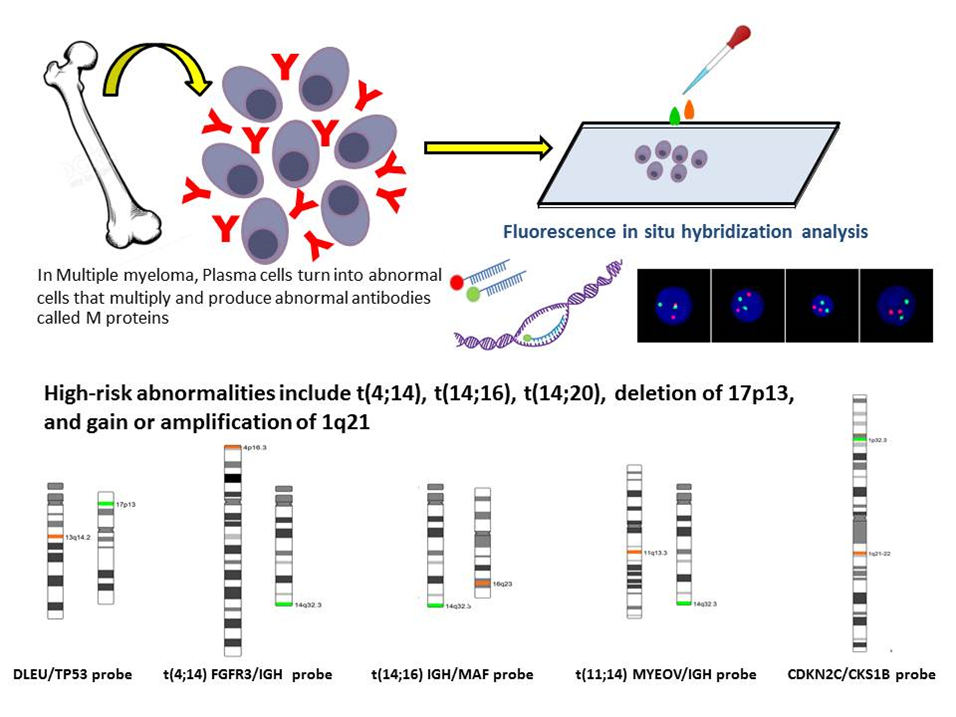
Background: Multiple myeloma (MM) is a genetically heterogeneous plasma cell malignancy with cytogenetic abnormalities that influence the prognosis and treatment outcomes. Fluorescence in situ hybridization (FISH) is crucial for detecting clinically significant abnormalities, including immunoglobulin heavy chain locus (IGH) translocations and deletions, e.g., del(17p), particularly in non-dividing plasma cells. However, cost and accessibility challenges limit comprehensive testing in resource-constrained settings, such as in Thailand.
Objective: This study aimed to investigate the incidence of cytogenetic abnormalities detected by FISH in MM cases over seven years in a Thai population, thereby highlighting regional trends and barriers to comprehensive testing.
Methods: A retrospective analysis was performed on 360 bone marrow samples from patients with MM between 2018 and 2024. FISH analysis targeted key abnormalities using specific probes: t(4;14), t(11;14), t(14;16), del(17p), and 1q21 amplification. Furthermore, the demographic data and testing frequencies were analyzed, as well as the prevalence rates of abnormalities were reported.
Results: Among the 360 cases, 47.4% exhibited abnormalities. The most common were del(17p) (30.6%), del(13q) (16.9%), and t(4;14) (6.9%). Testing limitations led to selective probe usage, with del (17p) probes ordered in 96.7% of cases, while 1q21 amplification probes were only ordered in 3.3% of cases. Regional trends revealed lower frequencies of t(11;14) compared to Western cohorts, suggesting ethnic influences.
Conclusion: FISH analysis revealed critical cytogenetic abnormalities in Thai patients with MM. However, financial constraints limit comprehensive testing, thus potentially hindering optimal risk stratification and treatment. Expanding diagnostic accessibility and integrating advanced technologies such as next-generation sequencing could address these barriers and improve patient outcomes.
Downloads
References
Clarke SE, Fuller KA, Erber WN. Chromosomal defects in multiple myeloma. Blood Rev 2024;64:101168.
https://doi.org/10.1016/j.blre.2024.101168
Malard F, Neri P, Bahlis NJ, Terpos E, Moukalled N, Hungria VTM, et al. Multiple myeloma. Nat Rev Dis Primers 2024;10:45.
https://doi.org/10.1038/s41572-024-00529-7
Abdallah N, Rajkumar SV, Greipp P, Kapoor P, Gertz MA, Dispenzieri A, et al. Cytogenetic abnormalities in multiple myeloma: association with disease characteristics and treatment response. Blood Cancer J 2020;10:82.
https://doi.org/10.1038/s41408-020-00348-5
Aydin C, Ulas T, Hangul C, Yucel OK, Iltar U, Salim O, et al. Conventional cytogenetics and interphase fluorescence in situ hybridization results in multiple myeloma: a Turkey Laboratory analysis of 381 cases. Indian J Hematol Blood Transfus 2020;36:284-91.
https://doi.org/10.1007/s12288-019-01215-5
Sonneveld P, Avet-Loiseau H, Lonial S, Usmani S, Siegel D, Anderson KC, et al. Treatment of multiple myeloma with high-risk cytogenetics: a consensus of the International Myeloma Working Group. Blood 2016;127:2955-62.
https://doi.org/10.1182/blood-2016-01-631200
Tian E. Fluorescence in situ hybridization (FISH) in multiple myeloma. Methods Mol Biol 2018;1792:55-69.
https://doi.org/10.1007/978-1-4939-7865-6_5
Kleber M, Ntanasis-Stathopoulos I, Terpos E. The Role of t(11;14) in tailoring treatment decisions in multiple myeloma. Cancers (Basel) 2023;15:5829.
https://doi.org/10.3390/cancers15245829
Umar M, Malik HS, Zaman B, Ahmad MW, Khan F, Nadeem H. Different translocations of multiple myeloma on fluorescent in situ hybridization (FISH) with clinical correlation. J Coll Physicians Surg Pak 2023;33:281-5.
https://doi.org/10.29271/jcpsp.2023.03.281
Yuan RF, Dong YJ, Li CR, Huang WR, Zhang LM, Zhu Q, et al. [Epidemiological analysis of cytogenetic abnormalities in patients with newly-diagnosed multiple myeloma: a multi-center retrospective study]. Zhonghua Xue Ye Xue Za Zhi 2020;41:10-5.
Ross FM, Avet-Loiseau H, Ameye G, Gutierrez NC, Liebisch P, O'Connor S, et al. Report from the European Myeloma Network on interphase FISH in multiple myeloma and related disorders. Haematologica 2012;97:1272-7.
https://doi.org/10.3324/haematol.2011.056176
Li S, Lim HH, Woo KS, Kim SH, Han JY. A retrospective analysis of cytogenetic alterations in patients with newly diagnosed multiple myeloma: a single center study in Korea. Blood Res 2016;51:122-6.
https://doi.org/10.5045/br.2016.51.2.122
Kim K, Lee JH, Kim JS, Min CK, Yoon SS, Shimizu K, et al. Clinical profiles of multiple myeloma in Asia-An Asian Myeloma Network study. Am J Hematol. 2014;89:751-6.
https://doi.org/10.1002/ajh.23731
Hanamura I. Gain/amplification of chromosome arm 1q21 in multiple myeloma. Cancers (Basel) 2021;13:256.
https://doi.org/10.3390/cancers13020256
Govindasamy P, Pandurangan P, Tarigopula A, Mani R, R Samuel C. Cytogenetic abnormalities in multiple myeloma patients at a tertiary healthcare center in India. Asian Pac J Cancer Prev 2019;20:235-41.
https://doi.org/10.31557/APJCP.2019.20.1.235
Hamdaoui H, Benlarroubia O, Ait Boujmia OK, Mossafa H, Ouldim K, Belkhayat A, et al. Cytogenetic and FISH analysis of 93 multiple myeloma Moroccan patients. Mol Genet Genomic Med 2020;8:e1363.
https://doi.org/10.1002/mgg3.1363
Huang SY, Yao M, Tang JL, Tsay W, Lee FY, Liu MC, et al. Clinical significance of cytogenetics and interphase fluorescence in situ hybridization analysis in newly diagnosed multiple myeloma in Taiwan. Ann Oncol 2005;16:1530-8.
https://doi.org/10.1093/annonc/mdi273
Sudha P, Ahsan A, Ashby C, Kausar T, Khera A, Kazeroun MH, et al. Myeloma genome project panel is a comprehensive targeted genomics panel for molecular profiling of patients with multiple myeloma. Clin Cancer Res 2022;28:2854-64.
Downloads
Published
How to Cite
Issue
Section
License
Copyright (c) 2025 Chulalongkorn Medical Journal

This work is licensed under a Creative Commons Attribution-NonCommercial-NoDerivatives 4.0 International License.


