
Dual role of Triphala extracts in breast cancer therapy: Enhanced paclitaxel cytotoxicity by ethanolic extract and cytoprotection by aqueous extract
Keywords:
Antioxidant therapy, breast cancer, chemotherapy, gallic acid, paclitaxel, reactive oxygen species, synergistic effect, triphalaAbstract
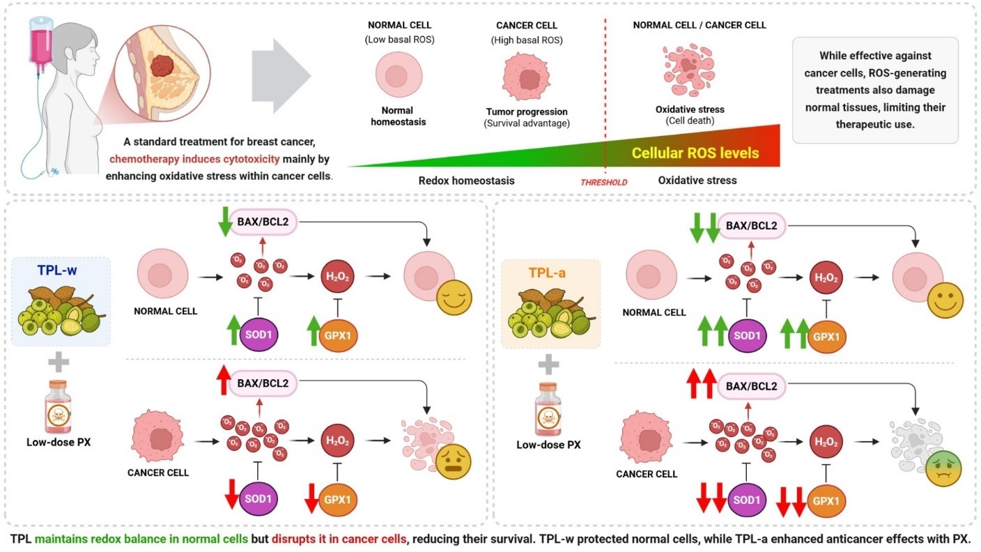
Background: Combining antioxidants with chemotherapy is an emerging strategy for reducing chemotoxicity in normal cells. Triphala (TPL), a polyphenol-rich Thai herbal formulation, has exhibited potential in enhancing chemotherapy efficacy; however, its interaction with paclitaxel in breast cancer treatment remains underexplored.
Objective: This study aimed to evaluate the effects of aqueous (TPL-w) and ethanolic (TPL-a) TPL extracts in combination with low-dose paclitaxel on breast cancer and non-tumorigenic mammary epithelial cells.
Methods: The bioactive components of TPL-w and TPL-a were identified using high-performance liquid chromatography. Thiazolyl blue tetrazolium bromide (MTT) and chloromethyl 22’ ,72’ - dichlorodihydrofluorescein diacetate (CM-H2DCFDA) assays were employed to assess cell viability and reactive oxygen species (ROS) levels, respectively, in breast cancer (MDA-MB-231, MCF-7) and epithelial (MCF-10A) cells. Combination index values were calculated using CompuSyn, whereas quantitative polymerase chain reaction was used to analyze apoptosis- and antioxidant-related gene expressions.
Results: Gallic acid was the predominant compound identified in both extracts, with higher levels detected in TPL-a. Paclitaxel and TPL-a cotreatment exhibited greater cytotoxicity in breast cancer cells, particularly in MDA-MB-231, compared with that of TPL-w cotreatment, primarily through enhanced ROS accumulation and an elevated BAX/BCL2 ratio, which led to apoptosis. TPL-a significantly downregulated superoxide dismutase (SOD1) and glutathione peroxidase (GPX1) expression in cancer cells, thereby enhancing oxidative stress. Conversely, TPL-w exhibited moderate cytotoxicity but effectively protected MCF-10A cells by upregulating the expression of SOD1 and GPX1, reducing ROS levels, and lowering the BAX/BCL2 ratio, thus enhancing cell survival.
Conclusion: TPL-a exhibited superior anticancer activity when combined with paclitaxel, whereas TPL-w provided stronger cytoprotection in normal cells. These findings highlight TPL-a’s potential as a complementary chemotherapeutic agent and TPL-w’s protective role, thereby warranting further investigation into their clinical applications.
Downloads
References
Giaquinto AN, Sung H, Newman LA, Freedman RA, Smith RA, Star J, et al. Breast cancer statistics 2024. CA Cancer J Clin 2024;74:477-95.
https://doi.org/10.3322/caac.21863
Perou CM, Sørlie T, Eisen MB, van de Rijn M, Jeffrey SS, Rees CA, et al. Molecular portraits of human breast tumours. Nature 2000;406:747-52.
https://doi.org/10.1038/35021093
Pham-Huy LA, He H, Pham-Huy C. Free radicals, antioxidants in disease and health. Int J Biomed Sci 2008;4:89-96.
https://doi.org/10.59566/IJBS.2008.4089
Yang HT, Villani RM, Wang HL, Simpson MJ, Roberts MS, Tang M, et al. The role of cellular reactive oxygen species in cancer chemotherapy. J Exp Clin Canc Res 2018;37:266.
https://doi.org/10.1186/s13046-018-0909-x
Weinberg F, Hamanaka R, Wheaton WW, Weinberg S, Joseph J, Lopez M, et al. Mitochondrial metabolism and ROS generation are essential for Kras-mediated tumorigenicity. Proc Natl Acad Sci U S A 2010;107:8788-93.
https://doi.org/10.1073/pnas.1003428107
Sarmiento-Salinas FL, Delgado-Magallón A, MontesAlvarado JB, Ramirez-Ramírez D, Flores-Alonso JC, Cortes-Hernandez P, et al. Breast cancer subtypes present a differential production of reactive oxygen species (ROS) and susceptibility to antioxidant treatment. Front Oncol 2019;9:480.
https://doi.org/10.3389/fonc.2019.00480
Charucharana S, Cheepsunthorn P, Cheepsunthorn CL. Antioxidant and chemotherapeutic synergy: Triphala enhances doxorubicin cytotoxicity in breast cancer cells and reduces toxicity in non-tumorigenic cells. Chula Med J 2025;69:95-108.
https://doi.org/10.56808/2673-060X.5560
Xiao H, Verdier-Pinard P, Fernandez-Fuentes N, Burd B, Angeletti R, Fiser A, et al. Insights into the mechanism of microtubule stabilization by Taxol. Proc Natl Acad Sci U S A 2006;103:10166-73.
https://doi.org/10.1073/pnas.0603704103
Yang HT, Villani RM, Wang HL, Simpson MJ, Roberts MS, Tang M, et al. The role of cellular reactive oxygen species in cancer chemotherapy. J Exp Clin Cancer Res 2018;37:266.
https://doi.org/10.1186/s13046-018-0909-x
Zhang Y, Tang Y, Tang X, Wang Y, Zhang Z, Yang H. Paclitaxel induces the apoptosis of prostate cancer cells via ROS-mediated HIF-1alpha expression. Molecules 2022;27:7183.
https://doi.org/10.3390/molecules27217183
Wargo JA, Reuben A, Cooper ZA, Oh KS, Sullivan RJ. Immune effects of chemotherapy, radiation, and targeted therapy and opportunities for combination with immunotherapy. Semin Oncol 2015;42:601 -16.
https://doi.org/10.1053/j.seminoncol.2015.05.007
Priyadrsini I, Mohan H, Naik G. An ayurvedic remedy - triphala as antioxidant drug. Antioxidants, Nutrients & Health. Free Radical Biol Med 2003;35 (Suppl):S43.
https://doi.org/10.1016/j.freeradbiomed.2003.09.027
Cheriyamundath S, Mahaddalkar T, Save SN, Choudhary S, Hosur RV, Lopus M. Aqueous extract of Triphala inhibits cancer cell proliferation through perturbation of microtubule assembly dynamics. Biomed Pharmacother 2018;98:76-81.
https://doi.org/10.1016/j.biopha.2017.12.022
Sandhya T, Mishra KP. Cytotoxic response of breast cancer cell lines, MCF 7 and T 47 D to triphala and its modification by antioxidants. Cancer Lett 2006;238: 304-13.
https://doi.org/10.1016/j.canlet.2005.07.013
Kaur S, Michael H, Arora S, Härkönen PL, Kumar S. The in vitro cytotoxic and apoptotic activity of Triphala - an Indian herbal drug. J Ethnopharmacol 2005;97:15-20.
https://doi.org/10.1016/j.jep.2004.09.050
Jagetia GC, Malagi KJ, Baliga MS, Venkatesh P, Veruva RR. Triphala, an ayurvedic Rasayana drug, protects mice against radiation-induced lethality by free-radical scavenging. J Altern Complement Med 2004;10:971-8.
https://doi.org/10.1089/acm.2004.10.971
Naik GH, Priyadarsini KI, Bhagirathi RG, Mishra B, Mishra KP, Banavalikar MM, et al. In vitro antioxidant studies and free radical reactions of triphala, an ayurvedic formulation and its constituents. Phytother Res 2005;19:582-6.
https://doi.org/10.1002/ptr.1515
Zhu J, Yi X, Zhang J, Chen S, Wu Y. Chemical profiling and antioxidant evaluation of Yangxinshi Tablet by HPLC-ESI-Q-TOF-MS/MS combined with DPPH assay. J Chromatogr B Analyt Technol Biomed Life Sci 2017;1060:262-71.
https://doi.org/10.1016/j.jchromb.2017.06.022
Meena AK, Narasimhaji CV, Velvizhi D, Singh A, Rekha P, Kumar V, et al. Determination of gallic acid in ayurvedic polyherbal formulation Triphala churna and its ingredients by HPLC and HPTLC. Res J Pharm Technol 2018;11:3243- 9.
https://doi.org/10.5958/0974-360X.2018.00596.6
Ainsworth EA, Gillespie KM. Estimation of total phenolic content and other oxidation substrates in plant tissues using Folin-Ciocalteu reagent. Nat Protoc 2007;2:875-7.
https://doi.org/10.1038/nprot.2007.102
Fattahi S, Zabihi E, Abedian Z, Pourbagher R, Motevalizadeh Ardekani A, Mostafazadeh A, et al. Total phenolic and flavonoid contents of aqueous extract of stinging nettle and in vitro antiproliferative effect on Hela and BT-474 cell lines. Int J Mol Cell Med 2014;3: 102-7.
Chou TC. Theoretical basis, experimental design, and computerized simulation of synergism and antagonism in drug combination studies. Pharmacol Rev 2006;58:621-81.
https://doi.org/10.1124/pr.58.3.10
Grace SC. Phenolics as antioxidants. In: Smirnoff N, editor. Antioxidants and reactive oxygen species in plants. Oxford, UK: Blackwell Publishing; 2005. p. 141-68.
https://doi.org/10.1002/9780470988565.ch6
Prasad S, Srivastava SK. Oxidative stress and cancer:chemopreventive and therapeutic role of Triphala.Antioxidants (Basel) 2020;9:72.
https://doi.org/10.3390/antiox9010072
Wang L, Weller CL. Recent advances in extraction of nutraceuticals from plants. Trends Food Sci Technol 2006;17:300-12.
https://doi.org/10.1016/j.tifs.2005.12.004
Panieri E, Santoro MM. ROS homeostasis and metabolism: a dangerous liason in cancer cells. Cell Death Dis 2016;7:e2253.
https://doi.org/10.1038/cddis.2016.105
Gorrini C, Harris IS, Mak TW. Modulation of oxidative stress as an anticancer strategy. Nat Rev Drug Discov 2013;12:931-47.
https://doi.org/10.1038/nrd4002
Abotaleb M, Liskova A, Kubatka P, Busselberg D. Therapeutic potential of plant phenolic acids in the treatment of cancer. Biomolecules 2020;10:221.
https://doi.org/10.3390/biom10020221
Sandhya T, Lathika KM, Pandey BN, Mishra KP. Potential of traditional ayurvedic formulation, Triphala, as a novel anticancer drug. Cancer Lett 2006;231:206-14.
https://doi.org/10.1016/j.canlet.2005.01.035
Èipák L, Novotný L, Èipáková I, Rauko P. Differential modulation of cisplatin and doxorubicin efficacies in leukemia cells by flavonoids. Nutr Res 2003;23:1045-57.
https://doi.org/10.1016/S0271-5317(03)00078-2
Ghatreh Samani K, Farrokhi E, Tabatabaee A, Jalilian N, Jafari M. Synergistic effects of lauryl gallate and tamoxifen on human breast cancer cell. Iran J Public Health 2020;49:1324-9.
https://doi.org/10.18502/ijph.v49i7.3586
Alexandre J, Batteux F, Nicco C, Chéreau C, Laurent A, Guillevin L, et al. Accumulation of hydrogen peroxide is an early and crucial step for paclitaxel-induced cancer cell death both in vitro and in vivo. Int J Cancer 2006;119:41-8.
https://doi.org/10.1002/ijc.21685
Moghtaderi H, Sepehri H, Delphi L, Attari F. Gallic acid and curcumin induce cytotoxicity and apoptosis in human breast cancer cell MDA-MB-231. Bioimpacts 2018;8:185-94.
https://doi.org/10.15171/bi.2018.21
Ighodaro OM, Akinloye OA. First line defence antioxidants-superoxide dismutase (SOD), catalase (CAT) and glutathione peroxidase (GPX): Their fundamental role in the entire antioxidant defence grid. Alex J Med 2018;54:287-93.
https://doi.org/10.1016/j.ajme.2017.09.001
Glorieux C, Zamocky M, Sandoval JM, Verrax J, Calderon PB. Regulation of catalase expression in healthy and cancerous cells. Free Radic Biol Med 2015;87:84-97.
https://doi.org/10.1016/j.freeradbiomed.2015.06.017
Walton PA, Brees C, Lismont C, Apanasets O, Fransen M. The peroxisomal import receptor PEX5 functions as a stress sensor, retaining catalase in the cytosol in times of oxidative stress. Biochim Biophys Acta Mol Cell Res 2017;1864:1833-43.
https://doi.org/10.1016/j.bbamcr.2017.07.013
Marklund SL, Westman NG, Lundgren E, Roos G. Copper- and zinc-containing superoxide dismutase, manganese-containing superoxide dismutase, catalase, and glutathione peroxidase in normal and neoplastic human cell lines and normal human tissues. Cancer Res 1982;42: 1955-61.
Wang S, Konorev EA, Kotamraju S, Joseph J, Kalivendi S, Kalyanaraman B. Doxorubicin induces apoptosis in normal and tumor cells via distinctly different mechanisms. intermediacy of H(2)O(2)- and p53-dependent pathways. J Biol Chem 2004;279:25535-43.
https://doi.org/10.1074/jbc.M400944200
Vijay K, Sowmya PR, Arathi BP, Shilpa S, Shwetha HJ, Raju M, et al. Low-dose doxorubicin with carotenoids selectively alters redox status and upregulates oxidative stress-mediated apoptosis in breast cancer cells. Food Chem Toxicol 2018;118:675-90.
Downloads
Published
How to Cite
Issue
Section
License
Copyright (c) 2025 Chulalongkorn Medical Journal

This work is licensed under a Creative Commons Attribution-NonCommercial-NoDerivatives 4.0 International License.


