
Periodontal status in patients with chronic skin diseases: a pilot study
Keywords:
Chronic skin disease, oral lesions, periodontal disease, periodontal statusAbstract
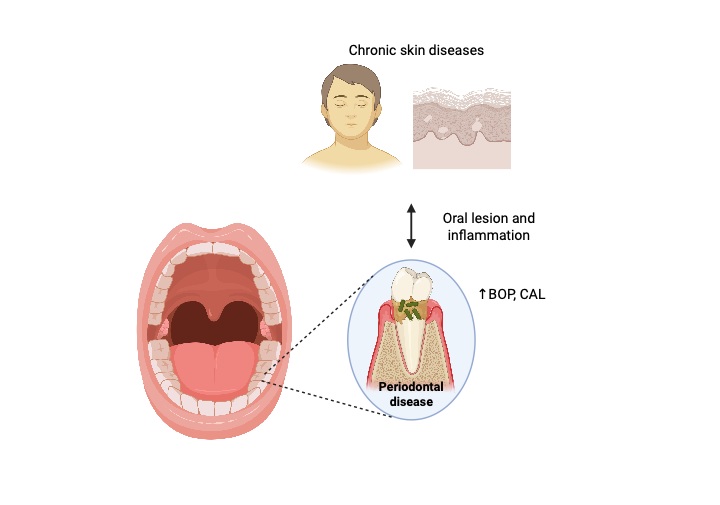
Background: Many skin diseases, especially those that also involve oral mucosa lesions, might play an essential role in the etiopathogenesis of oral health and mucocutaneous inflammation. The presence of these oral lesions may make it challenging to maintain satisfactory oral hygiene, which increases plaque accumulation and the risk of developing periodontal disease.
Objectives: This study evaluated and compared the periodontal status of patients with chronic skin disease (CSD) with that of healthy controls.
Methods: This study included 109 patients and 37 healthy controls. The evaluated parameters included the bleeding on probing index (BOP), periodontal pocket depths (PPD), clinical attachment level (CAL), simplified debris index (DI), simplified calculus index (CI), and the presence of oral lesions. Clinical parameters were measured and compared between the two groups using the chi-square test, t-test, and a non-parametric test.
Results: Patients with CSD had significantly higher BOP and a lower percentage of teeth with CAL d” 3 mm compared with those in the control group. Furthermore, CAL > 6 mm was only presented in the group of patients with CSD. There were no significant differences in the PPD, PI, and CI parameters and oral lesions between the two groups.
Conclusion: This pilot study revealed that the periodontal status of patients with CSD was worse than that of healthy controls. These results suggest that patients with CSD appear to be more at risk for the development and progression of periodontal diseases.
Downloads
References
Kiernan Y, O'Connor C, Ryan J, Murphy M. Oral health in patients with severe inflammatory dermatologic and rheumatologic disease. Skin Health Dis 2023;3:e156.
https://doi.org/10.1002/ski2.156
Akman A, Kacaroglu H, Yilmaz E, Alpsoy E. Periodontal status in patients with pemphigus vulgaris. Oral Dis 2008;14:640-3.
https://doi.org/10.1111/j.1601-0825.2008.01445.x
Arduino PG, Farci V, D'Aiuto F, Carcieri P, Carbone M,Tanteri C, et al. Periodontal status in oral mucous membrane pemphigoid: initial results of a case-control study. Oral Dis 2011;17:90-4.
https://doi.org/10.1111/j.1601-0825.2010.01709.x
Lo Russo L, Gallo C, Pellegrino G, Lo Muzio L, Pizzo G, Campisi G, et al. Periodontal clinical and microbiological data in desquamative gingivitis patients. Clin Oral Investig 2014;18:917-25.
https://doi.org/10.1007/s00784-013-1038-8
Lo Russo L, Guiglia R, Pizzo G, Fierro G, Ciavarella D, Lo Muzio L, et al. Effect of desquamative gingivitis on periodontal status: a pilot study. Oral Dis 2010;16:102-7.
https://doi.org/10.1111/j.1601-0825.2009.01617.x
Ramón-Fluixá C, Bagán-Sebastián J, Milián-Masanet M, Scully C. Periodontal status in patients with oral lichen planus: a study of 90 cases. Oral Dis 1999;5:303-6.
https://doi.org/10.1111/j.1601-0825.1999.tb00094.x
Schellinck AE, Rees TD, Plemons JM, Kessler HP,Rivera-Hidalgo F, Solomon ES. A comparison of the periodontal status in patients with mucous membrane pemphigoid: a 5-year follow-up. J Periodontol 2009;80:1765-73.
https://doi.org/10.1902/jop.2009.090244
Thorat MS, Raju A, Pradeep AR. Pemphigus vulgaris: effects on periodontal health. J Oral Sci 2010;52: 449-54.
https://doi.org/10.2334/josnusd.52.449
Tricamo MB, Rees TD, Hallmon WW, Wright JM, Cueva MA, Plemons JM. Periodontal status in patients with gingival mucous membrane pemphigoid. J Periodontol 2006;77:398-405.
https://doi.org/10.1902/jop.2006.050113
Jascholt I, Lai O, Zillikens D, Kasperkiewicz M. Periodontitis in oral pemphigus and pemphigoid: a systematic review of published studies. J Am Acad Dermatol 2017;76:975-8.e3.
https://doi.org/10.1016/j.jaad.2016.10.028
Macklis P, Adams K, Kaffenberger J, Kumar P, Krispinsky A, Kaffenberger B. The association between oral health and skin disease. J Clin Aesthet Dermatol 2020;13:48-53.
Rai NP, Kumar P, Mustafa SM, Divakar DD, Kheraif AA, Ramakrishnaiah R, et al. Relation between periodontal status and pre-cancerous condition (Oral Lichen Planus): a pilot study. Adv Clin Exp Med 2016;25:763-6.
https://doi.org/10.17219/acem/59014
Listgarten MA. Pathogenesis of periodontitis. J Clin Periodontol 1986;13:418-30.
https://doi.org/10.1111/j.1600-051X.1986.tb01485.x
Offenbacher S, Heasman PA, Collins JG. Modulation of host PGE2 secretion as a determinant of periodontal disease expression. J Periodontol 1993;64:432-44.
https://doi.org/10.1902/jop.1993.64.5s.432
Dhingra K, Vandana KL. Indices for measuring periodontitis: a literature review. Int Dent J 2011;61: 76-84.
https://doi.org/10.1111/j.1875-595X.2011.00018.x
Chapple ILC, Mealey BL, Van Dyke TE, Bartold PM,Dommisch H, Eickholz P, et al. Periodontal health and gingival diseases and conditions on an intact and a reduced periodontium: consensus report of workgroup 1 of the 2017 world workshop on the classification of periodontal and peri-implant diseases and conditions. J Periodontol 2018;89 Suppl 1:S74-S84.
Babay N, Alshehri F, Al Rowis R. Majors highlights of the new 2017 classification of periodontal and periimplant diseases and conditions. Saudi Dent J 2019;31:303-5.
https://doi.org/10.1016/j.sdentj.2019.04.006
Papapanou PN, Sanz M, Buduneli N, Dietrich T, Feres M, Fine DH, et al. Periodontitis: consensus report of workgroup 2 of the 2017 world workshop on the classification of periodontal and peri-Implant diseases and conditions. J Periodontol 2018;89 Suppl 1:S173-S82.
Greene JC, Vermillion JR. The simplified oral hygiene index. J Am Dent Assoc 1964;68:7-13.
Downloads
Published
How to Cite
Issue
Section
License
Copyright (c) 2025 Chulalongkorn Medical Journal

This work is licensed under a Creative Commons Attribution-NonCommercial-NoDerivatives 4.0 International License.


