
Comparison of radiation attenuation correction techniques for relative renal function calculation in renal cortical imaging
Keywords:
Attenuation correction, kidney depth, relative renal function, renal cortical imagingAbstract
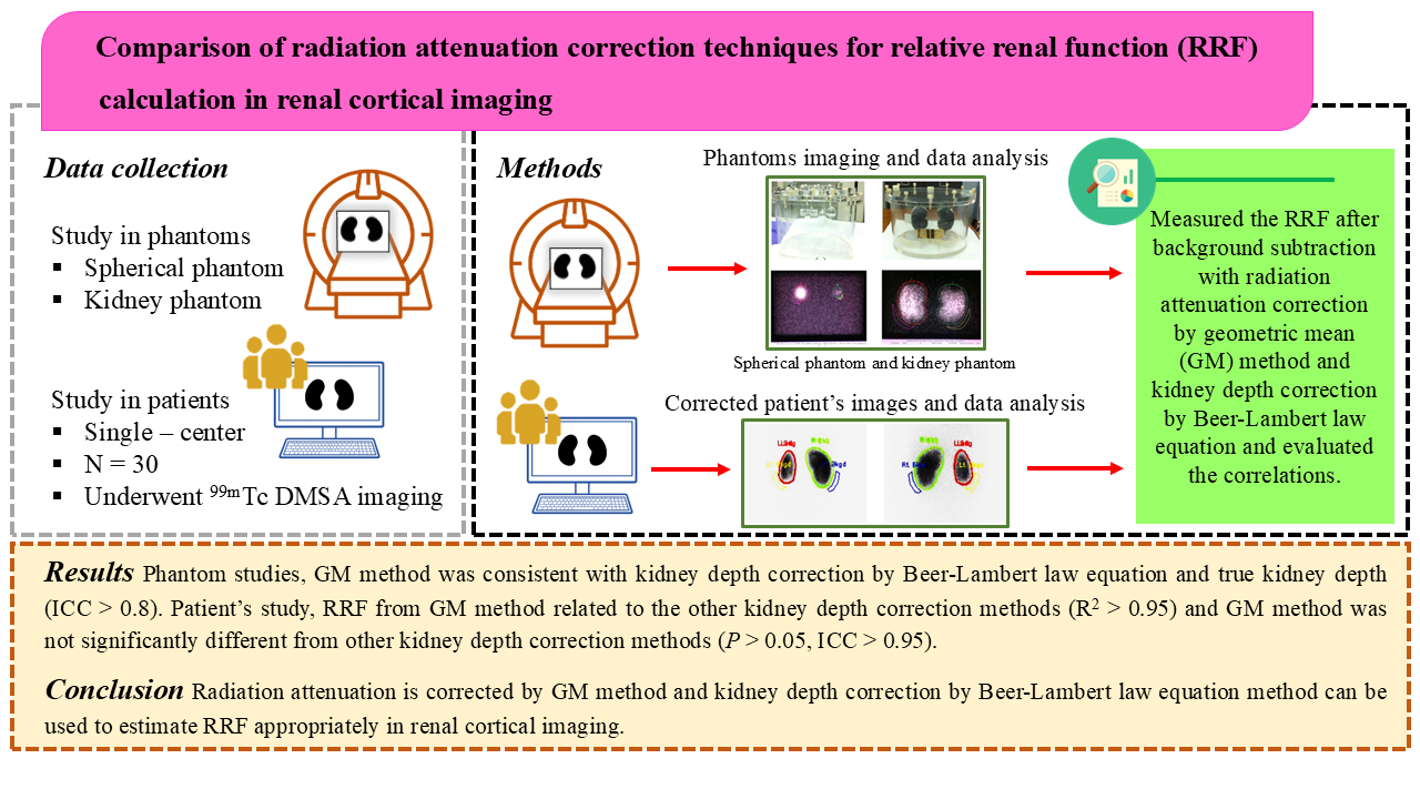
Background: A 99mTc-dimercaptosuccinic acid (99mTc-DMSA) scan is used for renal cortical imaging, specifically for the detection of renal cortical abnormalities, ectopic kidneys, renal scars, etc. Furthermore, relative renal function (RRF) is performed using 99mTc-DMSA planar imaging and estimated from the radiopharmaceutical uptake in the kidneys with background subtraction and radiation attenuation correction. Generally, attenuation correction is calculated from mathematical methods using the kidney depth correction (KDC) to obtain an accurate renal function value.
Objectives: This research aimed to study the correlation of the RRF after background subtraction with radiation attenuation correction using the geometric mean (GM) method and KDC according to the BeerLambert law equation.
Methods: Researchers studied the spherical phantom, the kidney phantom, and patients’ data. The phantom and patient data were acquired via gamma camera imaging. In all of the assessments, the background and kidney region of interest were created in planar images for subtracting the background counts and calculating the activity counts in the kidneys. This was used to record and analyze the data and then evaluate the RRF.
Results: For the RRF results with background subtraction and radiation attenuation correction in the phantom studies, the GM method was consistent with the KDC calculated using the Beer–Lambert law equation and the true kidney depth (intraclass correlation coefficient (ICC) > 0.8). The patient studies showed that the RRF results from the GM method correlated with the other KDC methods (R2 > 0.95), and the GM method was not significantly different from that of the other KDC calculation methods (P > 0.05, ICC > 0.95).
Conclusion: Radiation attenuation corrected with the GM method and KDC calculation using the Beer– Lambert law equation method can be used to appropriately estimate the RRF in renal cortical imaging.
Downloads
References
Marceau-Grimard M, Marion A, Côté C, Bolduc S, Dumont M, Moore K. Dimercaptosuccinic acid scintigraphy vs. ultrasound for renal parenchymal defects in children. Can Urol Assoc J 2017;11:260-4.
https://doi.org/10.5489/cuaj.4257
Piepsz A, Colarinha P, Gordon I, Hahn K, Olivier P, Roca I, et al. Guidelines for 99mTc- DMSA scintigraphy in children. Eur J Nucl Med 2001:28;BP37-41.
Lee KW, Bin KT, Jeong MS, Shong MH, Shin YT, Ro HK. Tc-99m dimercaptosuccinic acid (DMSA) renal scintigraphy in patients with acute pyelonephritis. Korean J Intern Med 1995;10:43-7.
https://doi.org/10.3904/kjim.1995.10.1.43
Vali R, Armstrong IS, Sever ZB, Biassoni L. SNMMI procedure standard/EANM practice guideline on pediatric [99mTc] Tc-DMSA renal cortical scintigraphy: an update. Clin Transl Imaging 2002:10;173-84.
https://doi.org/10.1007/s40336-022-00484-x
Guarino S, Capalbo D, Martin N, Campana G, Rambaldi PF, Miraglia Del Giudice E, et al. In children with urinary tract infection reduced kidney length and vesicoureteric reflux predict abnormal DMSA scan. Pediatr Res 2020;87:779-84.
https://doi.org/10.1038/s41390-019-0676-1
Lythgoe MF, Gradwell MJ, Evans K, Gordon I. Estimation and relevance of depth correction in paediatric renal studies. Eur J Nucl Med 1998;25:115-9.
https://doi.org/10.1007/s002590050202
Sontrapornpol T, Chaiwatanarat T, Kamklon N, Kawinthammasak C, Rattanamonrot R. Kidney depth calculation by anterior and posterior renal scintigraphy using attenuation - related techniques. Chula Med J 2017:61;425-38.
https://doi.org/10.58837/CHULA.CMJ.61.4.2
Xue J, Deng H, Jia X, Wang Y, Lu X, Ding X, et al. Establishing a new formula for estimating renal depth in a Chinese adult population. Medicine (Baltimore) 2017;96:e5940.
https://doi.org/10.1097/MD.0000000000005940
Koo TK, Li MY. A guideline of selecting and reporting intraclass correlation coefficients for reliability research. J Chiropr Med 2016;15:155-63.
https://doi.org/10.1016/j.jcm.2016.02.012
Chroustová D, Trnka J, Šírová V, Urbanová I, Langer J, Kubinyi J. Comparison of planar DMSA scan with an evaluation based on SPECT imaging in the split renal function assessment. Nucl Med Rev Cent East Eur 2016;19:12-7.
Downloads
Published
How to Cite
Issue
Section
License
Copyright (c) 2025 Chulalongkorn Medical Journal

This work is licensed under a Creative Commons Attribution-NonCommercial-NoDerivatives 4.0 International License.


