
Morphological and biological properties of platelet-rich fibrin membrane produced from different centrifugation protocols
Keywords:
Ocular surface diseases, platelet fibrin membrane, platelet-rich fibrin membraneAbstract
Background: Platelet-rich fibrin (PRF) membrane is a promising tool for the treatment of corneal epithelial disease; however, a standardized production protocol remains to be established.
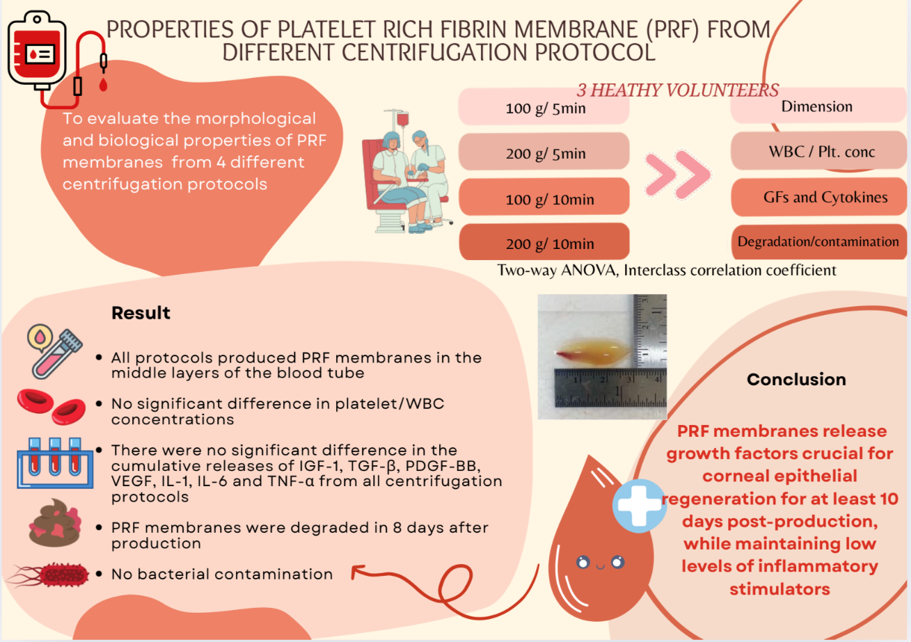
Objective: This study aimed to evaluate the morphological and biological properties of PRF membrane produced using different centrifugation protocols.
Methods: Four distinct low-speed centrifugation protocols (100 × g/5 min, 200 × g/5 min, 100 × g/10 min, and 200 × g/10 min) were established for PRF membrane production from blood obtained from three healthy volunteers. We assessed the fibrin and membrane dimensions, white blood cell and platelet concentrations, kinetic release of growth factors (TGF-b, PDGF-BB, VEGF, and IGF-1), and inflammatory cytokines (IL-6, IL-1, and TNF-a). Furthermore, in vitro degradation and bacterial contamination were examined.
Results: High platelet concentrations were consistently achieved in the PRF membranes produced by the four centrifugation protocols; however, there were no significant differences between these groups. Moreover, distinct release patterns were observed for each growth factor and cytokine. The PRF fibrin matrix effectively released growth factors over a sustained period of 3–10 days. Notably, no bacterial contamination was detected in any of the PRF membranes produced by the protocols.
Conclusion: Our findings definitively demonstrate that a low-speed centrifugation protocol can be employed to produce PRF membrane with high platelet concentrations that release growth factors over an extended period, thereby offering substantial therapeutic potential for ocular surface diseases.
Downloads
References
Giannaccare G, Versura P, Buzzi M, Primavera L,Pellagrini M, Compos EC. Blood derived eye drops for the treatment of cornea and ocular surface diseases.Transfus Apher Sci 2017;56:595-604.
https://doi.org/10.1016/j.transci.2017.07.023
Everts P, Onishi K, Jayaram P, Lana JF, Mautner K. Platelet-rich plasma: new performance understandings and therapeutic considerations in 2020. Int J Mol Sci 2020;21:7794.
https://doi.org/10.3390/ijms21207794
Dohan DM, Choukroun J, Diss A, Dohan SL, Dohan AJ, Mouhyi J, et al. Platelet-rich fibrin (PRF): a secondgeneration platelet concentrates. Part I: technological concepts and evolution. Oral Surg Oral Med Oral Pathol Oral Radiol Endod 2006;101:e37-44.
https://doi.org/10.1016/j.tripleo.2005.07.008
Can ME, Dereli Can G, Cagil N, Cakmak HB, Sungu N. Urgent therapeutic grafting of platelet-rich fibrin membrane in descemetocele. Cornea 2016;35:1245-9.
https://doi.org/10.1097/ICO.0000000000000917
Dohan DM, Choukroun J, Diss A, Dohan SL, Dohan AJ, Mouhyi J, et al. Platelet-rich fibrin (PRF): a secondgeneration platelet concentrate. Part III: leucocyte activation: a new feature for platelet concentrates? Oral Surg Oral Med Oral Pathol Oral Radiol Endod 2006;101:e51-5.
https://doi.org/10.1016/j.tripleo.2005.07.010
Miron R, Choukroun J, Ghanaati S. Controversies related to scientific report describing g-forces from studies on platelet-rich fibrin: Neccesity for standardization of relative centrifugation force values. Int J Growth Factors Stem Cell Dent 2018;1:80-9.
https://doi.org/10.4103/GFSC.GFSC_23_18
Masuki H, Okudera T, Watanebe T, Suzuki M, Nishiyama K, Okudera H, et al. Growth factor and proinflammatory cytokine contents in platelet-rich plasma (PRP), plasma rich in growth factors (PRGF), advanced platelet-rich fibrin (A-PRF), and concentrated growth factors (CGF). Int J Implant Dent 2016;2:19.
https://doi.org/10.1186/s40729-016-0052-4
Ghanaati S, Booms P, Orlowska A, Kubesch A, Lorenz J, Rutkowski J, et al. Advanced platelet-rich fibrin: a new concept for cell-based tissue engineering by means of inflammatory cells. J Oral Implantol 2014;40:679-89.
https://doi.org/10.1563/aaid-joi-D-14-00138
Dohan Ehrenfest M, Bielecki T, Jimbo R, Barbé G, Del Corso M, Inchingolo F, et al. Do the fibrin architecture and leukocyte content influence the growth factor release of platelet concentrates? An evidence-based answer comparing a pure platelet-rich plasma (P-PRP) gel and a leukocyte-and platelet-rich fibrin (L-PRF). Curr Pharm Biotechnol 2012;13:1145-52.
https://doi.org/10.2174/138920112800624382
Dohan Ehrenfest DM, Pinto NR, Pereda A, Jiménez P, Corso MD, Kang B-S, et al. The impact of the centrifuge characteristics and centrifugation protocols on the cells, growth factors, and fibrin architecture of a leukocyte-and platelet-rich fibrin (L-PRF) clot and membrane. Platelets 2018;29:171-84.
https://doi.org/10.1080/09537104.2017.1293812
Dohan Ehrenfest DM, de Peppo GM, Doglioli P, Sammartino G. Slow release of growth factors and thrombospondin-1 in Choukroun's platelet-rich fibrin (PRF): a gold standard to achieve for all surgical platelet concentrates technologies. Growth Factors 2009;27: 63-9.
https://doi.org/10.1080/08977190802636713
Kobayashi E, Flückiger L, Fujioka-Kobayashi M, Sawada K, Sculean A, Schaller B, et al. Comparative release of growth factors from PRP, PRF, and advancedPRF. Clin Oral Investig 2016;20:2353-60.
https://doi.org/10.1007/s00784-016-1719-1
Amable PR, Carias RBV, Teixeira MVT, da Cruz PachecoÍ, Corrêa do Amaral RJF, Granjeiro JM, et al. Plateletrich plasma preparation for regenerative medicine: optimization and quantification of cytokines and growth factors. Stem Cell Res Ther 2013;4:1-13.
https://doi.org/10.1186/scrt218
Pena JDdO, Melo GBd, Gomes JÁP, Haapalainen EF,Komagome CM, Santos NC, et al. Análise ultraestrutural e de fatores de crescimento de diferentes métodos de preservação da membrana amniótica utilizada em cirurgia ocular. Arq Bras Oftalmol 2007; 70:756-62.
https://doi.org/10.1590/S0004-27492007000500006
Koob TJ, Lim JJ, Zabek N, Massee M. Cytokines in single layer amnion allografts compared to multilayer amnion/chorion allografts for wound healing. J Biomed Mater Res B Appl Biomater 2015;103:1133-40.
https://doi.org/10.1002/jbm.b.33265
Bischoff M, Stachon T, Seitz B, Huber M, Zawada M, Langenbucher A, et al. Growth factor and interleukin concentrations in amniotic membrane-conditioned medium. Curr Eye Res 2017;42:174-80.
https://doi.org/10.3109/02713683.2016.1164189
Ding Z-Y, Tan Y, Peng Q, Zuo J, Li N. Novel applications of platelet concentrates in tissue regeneration. Exp Ther Med 2021;21:226.
https://doi.org/10.3892/etm.2021.9657
Sanders FWB, Huang J, Alió Del Barrio JL, Hamada S, McAlinden C. Amniotic membrane transplantation: structural and biological properties, tissue preparation, application, and clinical indications. Eye (Lond) 2024;38:668-79.
Downloads
Published
How to Cite
Issue
Section
License
Copyright (c) 2025 Chulalongkorn Medical Journal

This work is licensed under a Creative Commons Attribution-NonCommercial-NoDerivatives 4.0 International License.


