
Diagnostic accuracy of cerebral amyloid angiopathy criteria in the first pathologically confirmed Thai cohort: A pilot study
Keywords:
Cerebral amyloid angiopathy, diagnostic criteria, neuroimaging, neuropathologyAbstract
Background: The gold standard for diagnosing cerebral amyloid angiopathy (CAA) is full brain postmortem examination, which is rarely performed. Current diagnostic criteria are primarily based on clinical-radiological features and were developed from Western populations and thus may have limited applicability to Asian populations.
Objectives: We aimed to evaluate the accuracy of current diagnostic criteria and examine the clinicalradiological characteristics of Thai patients with CAA.
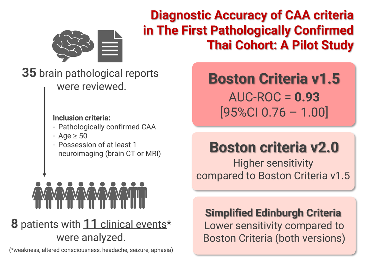
Methods: Brain histopathological specimens were reviewed from patients with clinical symptoms of CAA who underwent neurosurgical procedures, including intracerebral hemorrhage (ICH) evacuation, between 2011 and 2021 at King Chulalongkorn Memorial Hospital, Thai Red Cross Society. Patient characteristics and clinical events were collected retrospectively. Trained investigators systematically rated the radiological biomarkers from brain imaging performed closest to the date of pathological confirmation. The diagnostic accuracies of the Modified Boston Criteria v1.5, Boston Criteria v2.0, and Simplified Edinburgh Criteria were compared.
Results: Thirty-five pathological reports were reviewed. Eight patients (median age of 76.7 years) with confirmed CAA had 11 clinical events, including weakness, altered consciousness, headache, seizures, and aphasia. Receiver operating characteristic curve analysis revealed that the Boston Criteria v2.0 had higher sensitivity compared to the Modified Boston Criteria v1.5. Moreover, the Simplified Edinburgh Criteria demonstrated lower sensitivity compared to both of the Boston Criteria. The area under the curve for probable CAA using the Modified Boston Criteria v1.5 was 0.9 (95% confidence interval 0.8–1.0).
Conclusion: This pilot study reveals the diagnostic performance of CAA criteria and demonstrates its applicability among the Asian population. In resource-limited settings, the simplified Edinburgh criteria, which are computerized tomography-based criteria, are valuable for diagnosing patients with CAA-ICH. This is a pilot study with a relatively small sample size; larger studies with Asian cohorts are warranted to further validate these findings.
Downloads
References
Charidimou A, Boulouis G, Frosch MP, Baron JC, Pasi M, Albucher JF, et al. The Boston criteria version 2.0 for cerebral amyloid angiopathy: a multicentre, retrospective, MRI-neuropathology diagnostic accuracy study. Lancet Neurol 2022;21:714-25.
https://doi.org/10.1016/S1474-4422(22)00208-3
Charidimou A, Gang Q, Werring DJ. Sporadic cerebral amyloid angiopathy revisited: recent insights into pathophysiology and clinical spectrum. J Neurol Neurosurg Psychiatry 2012;83:124.
https://doi.org/10.1136/jnnp-2011-301308
Charidimou A, Boulouis G, Gurol ME, Ayata C, Bacskai BJ, Frosch MP, et al. Emerging concepts in sporadic cerebral amyloid angiopathy. Brain 2017;140:1829-50.
https://doi.org/10.1093/brain/awx047
Viswanathan A, Greenberg SM. Cerebral amyloid angiopathy in the elderly. Ann Neurol 2011;70:871-80.
https://doi.org/10.1002/ana.22516
Smith EE, Charidimou A, Ayata C, Werring DJ, Greenberg SM. Cerebral amyloid angiopathy-related transient focal neurologic episodes. Neurol 2021;97: 231-8.
https://doi.org/10.1212/WNL.0000000000012234
Greenberg SM, Edgar MA. Case records of the Massachusetts General Hospital. Weekly clinicopathological exercises. Case 22-1996. Cerebral hemorrhage in a 69-year-old woman receiving warfarin. N Engl J Med 1996;335:189-96.
https://doi.org/10.1056/NEJM199607183350308
Knudsen KA, Rosand J, Karluk D, Greenberg SM. Clinical diagnosis of cerebral amyloid angiopathy: validation of the Boston criteria. Neurol 2001;56:537-9.
https://doi.org/10.1212/WNL.56.4.537
Linn J, Halpin A, Demaerel P, Ruhland J, Giese AD, Dichgans M, et al. Prevalence of superficial siderosis in patients with cerebral amyloid angiopathy. Neurol 2010;74:1346-50.
https://doi.org/10.1212/WNL.0b013e3181dad605
Sembill JA, Knott M, Xu M, Roeder SS, Hagen M, Sprügel MI, et al. Simplified Edinburgh CT criteria for identification of lobar intracerebral hemorrhage associated with cerebral amyloid angiopathy. Neurol 2022;98:e1997-2004.
https://doi.org/10.1212/WNL.0000000000200261
Rodrigues MA, Samarasekera N, Lerpiniere C, Humphreys C, McCarron MO, White PM, et al. The Edinburgh CT and genetic diagnostic criteria for lobar intracerebral haemorrhage associated with cerebral amyloid angiopathy: model development and diagnostic test accuracy study. Lancet Neurol 2018; 17:232-40.
https://doi.org/10.1016/S1474-4422(18)30006-1
Schwarz G, Banerjee G, Hostettler IC, Ambler G, Seiffge DJ, Ozkan H, et al. MRI and CT imaging biomarkers of cerebral amyloid angiopathy in lobar intracerebral hemorrhage. Inter J Stroke 2023;18:85-94.
https://doi.org/10.1177/17474930211062478
Greenberg SM, Charidimou A. Diagnosis of cerebral amyloid angiopathy. Stroke 2018;49:491-7.
https://doi.org/10.1161/STROKEAHA.117.016990
Vonsattel JP, Myers RH, Hedley-Whyte ET, Ropper AH, Bird ED, Richardson EP, Jr. Cerebral amyloid angiopathy without and with cerebral hemorrhages: a comparative histological study. Ann Neurol 1991;30: 637-49.
https://doi.org/10.1002/ana.410300503
Greenberg SM, Vonsattel JP. Diagnosis of cerebral amyloid angiopathy. Sensitivity and specificity of cortical biopsy. Stroke 1997;28:1418-22.
https://doi.org/10.1161/01.STR.28.7.1418
Duering M, Biessels GJ, Brodtmann A, Chen C, Cordonnier C, de Leeuw FE, et al. Neuroimaging standards for research into small vessel diseaseadvances since 2013. Lancet Neurol 2023;22:602-18.
https://doi.org/10.1016/S1474-4422(23)00131-X
Charidimou A, Linn J, Vernooij MW, Opherk C, Akoudad S, Baron J-C, et al. Cortical superficial siderosis:detection and clinical significance in cerebral amyloid angiopathy and related conditions. Brain 2015;138:2126-39.
https://doi.org/10.1093/brain/awv162
Charidimou A, Frosch MP, Al-Shahi Salman R, BaronJC, Cordonnier C, Hernandez-Guillamon M, et al. Advancing diagnostic criteria for sporadic cerebral amyloid angiopathy: study protocol for a multicenter MRI-pathology validation of Boston criteria v2.0. Int J Stroke 2019;14:956-71.
https://doi.org/10.1177/1747493019855888
von Elm E, Altman DG, Egger M, Pocock SJ, Gøtzsche PC, Vandenbroucke JP. Strengthening the Reporting of Observational Studies in Epidemiology (STROBE) statement: guidelines for reporting observational studies. BMJ 2007;335:806-8.
https://doi.org/10.1136/bmj.39335.541782.AD
Puy L, Pasi M, Rodrigues M, van Veluw SJ, Tsivgoulis G, Shoamanesh A, et al. Cerebral microbleeds: from depiction to interpretation. J Neurol Neurosurg Psychiatry 2021;92:598.
https://doi.org/10.1136/jnnp-2020-323951
Conijn MM, Geerlings MI, Biessels GJ, Takahara T, Witkamp TD, Zwanenburg JJ, et al. Cerebral microbleeds on MR imaging: comparison between 1.5 and 7T. AJNR Am J Neuroradiol 2011;32:1043-9.
https://doi.org/10.3174/ajnr.A2450
Cordonnier C, Klijn C, Smith EE, Al-Shahi Salman R, Chwalisz BK, van Etten E, et al. Diagnosis and management of cerebral amyloid angiopathy: a scientific statement from the International CAA Association and the World Stroke Organization. Int J Stroke 2025: 17474930251365861.
https://doi.org/10.1177/17474930251365861
Jäkel L, De Kort AM, Klijn CJM, Schreuder F, Verbeek MM. Prevalence of cerebral amyloid angiopathy: a systematic review and meta-analysis. Alzheimers Dement 2022;18:10-28.
https://doi.org/10.1002/alz.12366
van Etten ES, Kaushik K, van Zwet EW, Voigt S, van Walderveen MAA, van Buchem MA, et al. Sensitivity of the Edinburgh criteria for lobar intracerebral hemorrhage in hereditary cerebral amyloid angiopathy. Stroke 2020;51:3608-12.
https://doi.org/10.1161/STROKEAHA.120.031264
Hostettler IC, Wilson D, Fiebelkorn CA, Aum D, Ameriso SF, Eberbach F, et al. Risk of intracranial haemorrhage and ischaemic stroke after convexity subarachnoid haemorrhage in cerebral amyloid angiopathy: international individual patient data pooled analysis. J Neurol 2022;269:1427-38.
https://doi.org/10.1007/s00415-021-10706-3
Charidimou A, Boulouis G, Greenberg SM, Viswanathan A. Cortical superficial siderosis and bleeding risk in cerebral amyloid angiopathy: a meta-analysis. Neurol 2019;93:e2192-e202.
https://doi.org/10.1212/WNL.0000000000008590
Charidimou A, Boulouis G, Roongpiboonsopit D, Xiong L, Pasi M, Schwab KM, et al. Cortical superficial siderosis and recurrent intracerebral hemorrhage risk in cerebral amyloid angiopathy: large prospective cohort and preliminary meta-analysis. Int J Stroke 2019;14:723-33.
https://doi.org/10.1177/1747493019830065
Charidimou A, Imaizumi T, Moulin S, Biffi A, Samarasekera N, Yakushiji Y, et al. Brain hemorrhage recurrence, small vessel disease type, and cerebral microbleeds: a meta-analysis. Neurol 2017;89:820-9.
https://doi.org/10.1212/WNL.0000000000004259
Charidimou A, Boulouis G, Xiong L, Pasi M, Roongpiboonsopit D, Ayres A, et al. Cortical superficial siderosis evolution. Stroke 2019;50:954-62.
https://doi.org/10.1161/STROKEAHA.118.023368
Pongpitakmetha T, Fotiadis P, Pasi M, Boulouis G, Xiong L, Warren AD, et al. Cortical superficial siderosis progression in cerebral amyloid angiopathy: prospective MRI study. Neurol 2020;94:e1853-65.
https://doi.org/10.1212/WNL.0000000000009321
Pasi M, Sugita L, Xiong L, Charidimou A, Boulouis G, Pongpitakmetha T, et al. Association of cerebral small vessel disease and cognitive decline after intracerebral hemorrhage. Neurol 2021;96:e182-92.
https://doi.org/10.1212/WNL.0000000000011050
Xiong L, Charidimou A, Pasi M, Boulouis G, Pongpitakmetha T, Schirmer MD, et al. Predictors for late post-intracerebral hemorrhage dementia in patients with probable cerebral amyloid angiopathy. J Alzheimers Dis 2019;71:435-42.
https://doi.org/10.3233/JAD-190346
Greenberg SM, Ziai WC, Cordonnier C, Dowlatshahi D, Francis B, Goldstein JN, et al. 2022 guideline for the management of patients with spontaneous intracerebral hemorrhage: a guideline from the American Heart Association/American Stroke Association. Stroke 2022;53:e282-e361.
https://doi.org/10.1161/STR.0000000000000407
Greenberg SM, Aparicio HJ, Furie KL, Goyal MS, Hinman JD, Kozberg M, et al. Vascular neurology considerations for antiamyloid immunotherapy: a science advisory from the American Heart Association. Stroke 2025;56:e30-e8.
https://doi.org/10.1161/STR.0000000000000480
Greenberg SM, Cordonnier C, Schneider JA, Smith EE, van Buchem MA, van Veluw SJ, et al. Off-label use of aducanumab for cerebral amyloid angiopathy. Lancet Neurol 2021;20:596-7.
https://doi.org/10.1016/S1474-4422(21)00213-1
Koduri S, Keep RF, Xi G, Chaudhary N, Pandey AS. The role of iron in hemorrhagic stroke. Stroke Vasc Interv Neurol 2022;2:e000419.
https://doi.org/10.1161/SVIN.122.000419
Magid-Bernstein J, Girard R, Polster S, Srinath A, Romanos S, Awad IA, et al. Cerebral hemorrhage: pathophysiology, treatment, and future directions. Circ Res 2022;130:1204-29.
https://doi.org/10.1161/CIRCRESAHA.121.319949
Wei Y, Song X, Gao Y, Gao Y, Li Y, Gu L. Iron toxicity in intracerebral hemorrhage: physiopathological and therapeutic implications. Brain Res Bull 2022;178: 144-54.
https://doi.org/10.1016/j.brainresbull.2021.11.014
Pongpitakmetha T, Thanprasertsuk S. The diagnostic accuracy of cerebral amyloid angiopathy criteria between modified Boston criteria v1.5, Boston criteria v2.0, and simplified Edinburgh criteria - the first Thai cerebral amyloid angiopathy pathological confirmed cohort. Alzheimer's Dement 2023;19:e076706.
Downloads
Published
How to Cite
Issue
Section
License
Copyright (c) 2026 Chulalongkorn Medical Journal

This work is licensed under a Creative Commons Attribution-NonCommercial-NoDerivatives 4.0 International License.


