
Normal appendiceal diameter on computed tomography and its influencing factors in the pediatric population
Keywords:
Appendicitis, appendix, computed tomography (CT), normal childrenAbstract
Background: Appendicitis is the most common indication for emergency abdominal surgery in children. Computed tomography (CT) plays a crucial role in evaluating pediatric patients with suspected appendicitis. However, the literature describing the normal appendiceal diameter in the pediatric Asian population remains limited.
Objectives: This study aimed to establish reference values for normal appendiceal diameter and to identify variables influencing its diameter in pediatric patients aged 18 years or younger who underwent abdominal CT.
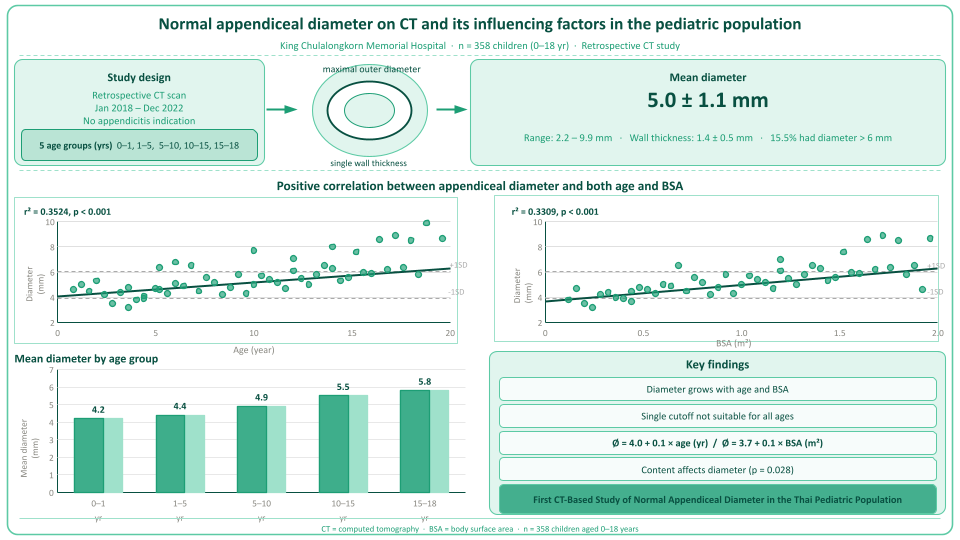
Methods: The study was reviewed and approved by the Institutional Review Board. We conducted a retrospective study of 358 children (≤18 years of age) who underwent abdominal CT scans between January 1, 2018, and December 31, 2022, for indications unrelated to appendicitis. Demographic data, including calculated body mass index and body surface area (BSA), were recorded. Radiologic data on the appendices included the maximal outer diameter and single-wall thickness. The data were classified into five age-based strata, and the mean appendiceal diameters were calculated. Associations between appendiceal diameter and age, and between appendiceal diameter and BSA, were assessed using linear regression models.
Results: The mean appendiceal diameter was 5.0 ± 1.1 mm. Stratified analysis by age revealed the following mean appendiceal diameters: 4.2 mm (0–1 years), 4.4 mm (1–5 years), 4.9 mm (5–10 years), 5.5 mm (10–15 years), and 5.8 mm (15–18 years). Age and BSA demonstrated significant predictive variability for appendiceal diameter (P < 0.001), explaining 35.2% and 33.1% of the variance, respectively. The regression equations were: appendiceal diameter (mm) = 4.0 + 0.1 × age (years) and appendiceal diameter (mm) = 3.7 + 0.1 × BSA (m2).
Conclusion: A uniform diameter cutoff for appendiceal diameter should not be applied across the pediatric population because the appendix grows during childhood. This study demonstrated positive correlations between the appendiceal diameter and age and between the appendiceal diameter and BSA.
Downloads
References
Becker C, Kharbanda A. Acute appendicitis in pediatric patients: an evidence-based review. Pediatr Emerg Med Pract 2019;16:1-20.
Koberlein GC, Trout AT, Rigsby CK, Iyer RS, Alazraki AL, Anupindi SA, et al. ACR Appropriateness Criteria(®) Suspected Appendicitis-Child. J Am Coll Radiol 2019;16:S252-S63.
https://doi.org/10.1016/j.jacr.2019.02.022
Strouse PJ. Pediatric appendicitis: an argument for US. Radiology 2010;255:8-13.
https://doi.org/10.1148/radiol.10091198
Callahan MJ, Rodriguez DP, Taylor GA. CT of appendicitis in children. Radiology. 2002;224:325-32.
https://doi.org/10.1148/radiol.2242010998
Taylor GA, Callahan MJ, Rodriguez D, Smink DS. CT for suspected appendicitis in children: an analysis of diagnostic errors. Pediatr Radiol 2006;36:331-7.
https://doi.org/10.1007/s00247-005-0079-9
Swenson DW, Schooler GR, Stamoulis C, Lee EY. MRI of the normal appendix in children: data toward a new reference standard. Pediatr Radiol 2016;46:1003-10.
https://doi.org/10.1007/s00247-016-3559-1
To T, Langer JC. Does access to care affect outcomes of appendicitis in children?--A population-based cohort study. BMC Health Serv Res 2010;10:250.
https://doi.org/10.1186/1472-6963-10-250
Rao PM, Rhea JT, Rattner DW, Venus LG, Novelline RA. Introduction of appendiceal CT: impact on negative appendectomy and appendiceal perforation rates. Ann Surg 1999;229:344-9.
https://doi.org/10.1097/00000658-199903000-00007
Oyetunji TA, Ong'uti SK, Bolorunduro OB, Cornwell EE 3rd, Nwomeh BC. Pediatric negative appendectomy rate: trend, predictors, and differentials. J Surg Res 2012;173:16-20.
https://doi.org/10.1016/j.jss.2011.04.046
Bachur RG, Hennelly K, Callahan MJ, Chen C, Monuteaux MC. Diagnostic imaging and negative appendectomy rates in children: effects of age and gender. Pediatrics 2012;129:877-84.
https://doi.org/10.1542/peds.2011-3375
Karabulut R, Sonmez K, Turkyilmaz Z, Demirogullari B, Ozen IO, Demirtola A, et al. Negative appendectomy experience in children. Ir J Med Sci. 2011;180:55-8.
https://doi.org/10.1007/s11845-010-0526-y
Trout AT, Towbin AJ, Zhang B. Journal club: The pediatric appendix: defining normal. AJR Am J Roentgenol. 2014;202:936-45.
https://doi.org/10.2214/AJR.13.11030
Sivit CJ, Siegel MJ, Applegate KE, Newman KD. When appendicitis is suspected in children. Radiographics 2001;21:247-62.
https://doi.org/10.1148/radiographics.21.1.g01ja17247
Ozturkmen Akay H, Akpinar E, Akgul Ozmen C, Ergun O, Haliloglu M. Visualization of the normal appendix in children by non-contrast MDCT. Acta Chir Belg 2007;107:531-4.
https://doi.org/10.1080/00015458.2007.11680116
Orscheln ES, Trout AT. Appendiceal diameter: CT versus sonographic measurements. Pediatr Radiol 2016;46:316-21.
https://doi.org/10.1007/s00247-015-3491-9
Trout AT, Zhang B, Towbin AJ. Measurement error in CT assessment of appendix diameter. Pediatr Radiol 2016;46:1831-6.
https://doi.org/10.1007/s00247-016-3699-3
Lowe LH, Penney MW, Scheker LE, Perez R Jr, Stein SM, Heller RM, et al. Appendicolith revealed on CT in children with suspected appendicitis: how specific is it in the diagnosis of appendicitis? AJR Am J Roentgenol 2000;175:981-4.
https://doi.org/10.2214/ajr.175.4.1750981
Krishnamoorthi R, Ramarajan N, Wang NE, Newman B, Rubesova E, Mueller CM, et al. Effectiveness of a staged US and CT protocol for the diagnosis of pediatric appendicitis: reducing radiation exposure in the age of ALARA. Radiology 2011;259:231-9.
https://doi.org/10.1148/radiol.10100984
Wiersma F, Srámek A, Holscher HC. US features of the normal appendix and surrounding area in children. Radiology 2005;235:1018-22.
https://doi.org/10.1148/radiol.2353040086
Ozel A, Orhan UP, Akdana B, Disli C, Erturk SM, Basak M, et al. Sonographic appearance of the normal appendix in children. J Clin Ultrasound 2011;39:183-6.
https://doi.org/10.1002/jcu.20807
Queiroz MRG, Francisco Neto MJ, Rahal Junior A, Jabour VA, Andrade GNL, Silva PSDD, et al. Ultrasonographic evaluation of cecal appendix diameter in pediatric population. Einstein (Sao Paulo) 2022;20:eAO6935.
https://doi.org/10.31744/einstein_journal/2022AO6935
Fukuta A, Kakiuchi T, Sadashima E, Inoue T, Muramori K. Reference growth curves for normal appendiceal diameter in childhood. Sci Rep 2020;10:12206.
Downloads
Published
How to Cite
Issue
Section
License
Copyright (c) 2026 Chulalongkorn Medical Journal

This work is licensed under a Creative Commons Attribution-NonCommercial-NoDerivatives 4.0 International License.


