
Pituitary apoplexy in a 19-year-old woman: An uncommon age and gender presentation
Keywords:
Bilateral hemianopia, case reports, optic chiasm, pituitary adenoma, pituitary apoplexyAbstract
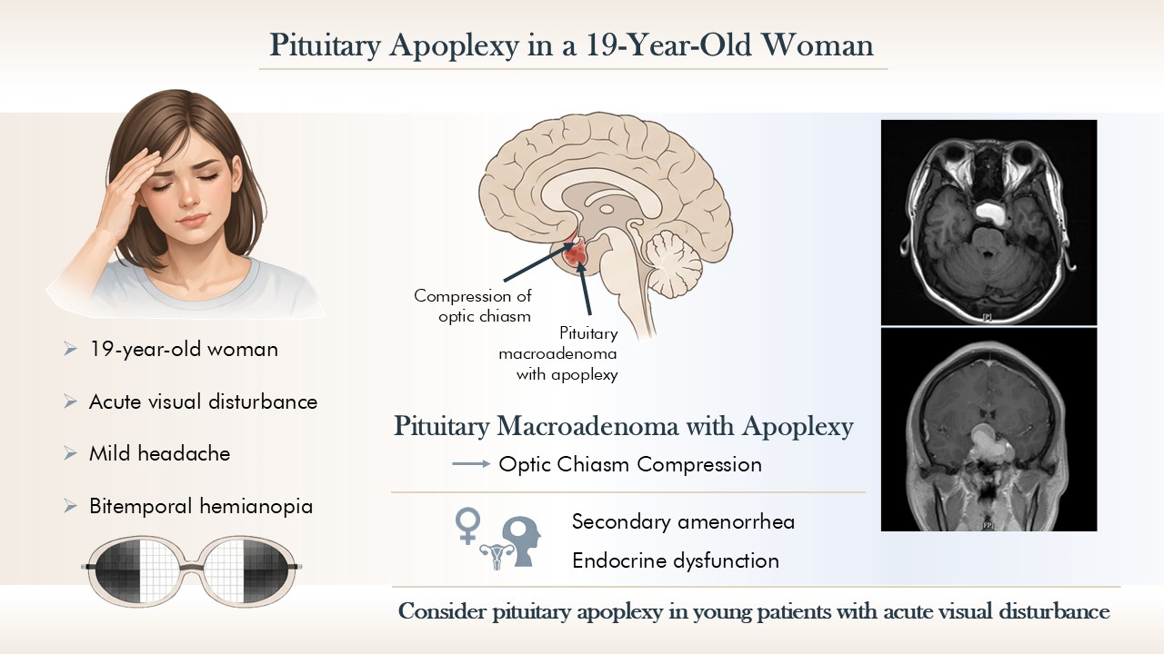
Pituitary apoplexy is a rare clinical syndrome that is caused by acute hemorrhage or infarction of the pituitary gland. It is most frequently reported in men during the 5thand 6thdecades of life and is rarely encountered in young women. In this report, we describe a 19-year-old woman who presented with acute visual disturbance accompanied by occasional mild headaches. Ophthalmologic examination revealed bitemporal hemianopia, and head magnetic resonance imaging demonstrated an intrasellar mass with suprasellar extension, intratumoral hemorrhage, and optic chiasm compression, consistent with a pituitary macroadenoma with apoplexy. The patient also had a history of secondary amenorrhea, thereby suggesting possible endocrine dysfunction. This case highlights the importance of considering pituitary apoplexy in young patients with acute visual disturbances.
Downloads
References
Iglesias, P. Pituitary apoplexy: An updated review. J Clin Med 2024;13:2508.
https://doi.org/10.3390/jcm13092508
Boellis A, di Napoli A, Romano A, Bozzao A. Pituitary apoplexy: an update on clinical and imaging features. Insights Imaging 2014;5:753-62.
https://doi.org/10.1007/s13244-014-0362-0
Rajasekaran S, Vanderpump M, Baldeweg S, Drake W, Reddy N, Lanyon M, et al. UK guidelines for the management of pituitary apoplexy. Clin Endocrinol (Oxf) 2011;74:9-20.
https://doi.org/10.1111/j.1365-2265.2010.03913.x
Donegan D, Erickson D. Revisiting pituitary apoplexy. J Endocr Soc 2022;6:bvac113.
https://doi.org/10.1210/jendso/bvac113
Hadj Kacem F, Trimeche O, Gargouri I, Ben Salah D, Charfi N, Rekik N, et al. Diagnosis and management of pituitary apoplexy: a Tunisian data. Chin Neurosurg J 2023;9:17.
https://doi.org/10.1186/s41016-023-00331-6
Falhammar H, Tornvall S, Höybye C. Pituitary apoplexy: A retrospective study of 33 cases from a single center. Front Endocrinol (Lausanne) 2021;12:656950.
https://doi.org/10.3389/fendo.2021.656950
Moscona-Nissan A, Sidauy-Adissi J, Hermoso-Mier KX, Glick-Betech SS, Chávez-Vera LJ, Martinez-Mendoza F, et al. Diagnosis and treatment of pituitary apoplexy, A true endocrine emergency. Arch Med Res 2024;55:103001.
https://doi.org/10.1016/j.arcmed.2024.103001
Fernandez A, Karavitaki N, Wass JAH. Prevalence of pituitary adenomas: A community-based, cross-sectional study in Banbury (Oxfordshire, UK). Clin Endocrinol 2010;72:377-82.
https://doi.org/10.1111/j.1365-2265.2009.03667.x
Raappana A, Koivukangas J, Ebeling T, Pirilä T. Incidence of pituitary adenomas in Northern Finland in 1992-2007. J Clin Endocrinol Metab 2010;95:4268-75.
https://doi.org/10.1210/jc.2010-0537
Briet C, Salenave S, Bonneville JF, Laws ER, Chanson P. Pituitary apoplexy. Endocr Rev 2015;36:622-45.
https://doi.org/10.1210/er.2015-1042
Muthukumar N. Pituitary apoplexy: A comprehensive review. Neurol India 2020;68 Supplement:S72-S8.
https://doi.org/10.4103/0028-3886.287669
Sakalas R, David RB, Vines FS, Becker DP. Pituitary apoplexy in a child: Case report. J Neurosurg 1973;39:519-22.
https://doi.org/10.3171/jns.1973.39.4.0519
Ohata H, Shinda B, Nakamura H, Teranishi Y, Morisako H, Ohata K, et al. Pituitary apoplexy in a puberal child: A case report and review of the literature. Interdiscip Neurosurg 2021;25:101160.
https://doi.org/10.1016/j.inat.2021.101160
Bonneville F, Cattin F, Marsot-Dupuch K, Dormont D, Bonneville JF, Chiras J. T1 signal hyperintensity in the sellar region: spectrum of findings. Radiographics 2006;26:93-113.
https://doi.org/10.1148/rg.261055045
Banskota S, Adamson DC. Pituitary adenomas: From diagnosis to therapeutics.Biomedicines 2021;9:494.
https://doi.org/10.3390/biomedicines9050494
Molitch ME. Diagnosis and treatment of pituitary adenomas: A review. JAMA 2017;317:516-24.
https://doi.org/10.1001/jama.2016.19699
Qin J, Li K, Wang X, Bao Y. A comparative study of functioning and non-functioning pituitary adenomas. Medicine (Baltimore) 2021;100:e25306.
https://doi.org/10.1097/MD.0000000000025306
Fleseriu M, Karavitaki N. Non-functioning pituitary adenomas, not all the same and certainly not boring! Pituitary 2018;21:109-10.
https://doi.org/10.1007/s11102-018-0875-5
Haidenberg-David F, Sidauy-Adissi J, Moscona-Nissan A, Jonguitud-Zumaya E, Fugarolas-Morinelli M, Martinez-Mendoza F, et al. Overview of hyperprolactinemia:general approach and reproductive health implications. Arch Med Res 2024;55:103102.
https://doi.org/10.1016/j.arcmed.2024.103102
Melmed S, Casanueva FF, Hoffman AR, Kleinberg DL, Montori VM, Schlechte JA, Wass JA. Diagnosis and treatment of hyperprolactinemia: An endocrine society clinical practice guideline. J Clin Endocrinol Metab 2011;96:273-88.
https://doi.org/10.1210/jc.2010-1692
American Academy of Ophthalmology. 2025-2026 Basic and clinical science course section 5: Neuro-Ophthalmology. San Francisco (CA): American Academy of Ophthalmology; 2025.
Singh V, Holmes R. Visual recovery following surgical intervention for pituitary apoplexy correlated with preoperative optical coherence tomography. N Z Med J 2022;135:122-9.
Downloads
Published
How to Cite
Issue
Section
License
Copyright (c) 2026 Chulalongkorn Medical Journal

This work is licensed under a Creative Commons Attribution-NonCommercial-NoDerivatives 4.0 International License.


