
Incidental leftward displacement of the appendix due to redundant sigmoid colon: A case report of anatomical variation
Keywords:
Anatomical variation, appendix, case report, redundant colonAbstract
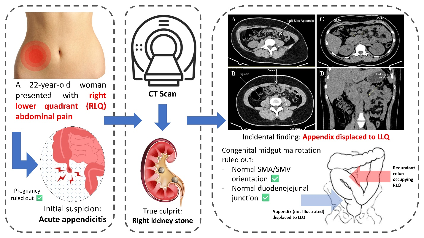
A left-sided appendix is an uncommon clinical finding, often attributed to congenital anomalies, such as situs inversus or intestinal malrotation. However, it can also arise from anatomical displacement by other organs, such as redundant colonic loops. While rare, such variations may complicate the clinical and radiological diagnosis of abdominal pain and result in potentially serious consequences if not recognized. This case report highlights a left-sided appendix due to a redundant sigmoid colon incidentally discovered during the evaluation of right lower quadrant pain. A 22-year-old woman presented to the emergency department with right flank pain, which later localized to the right lower quadrant (RLQ). There was localized tenderness in the RLQ, with no systemic symptoms. A working diagnosis of acute appendicitis was considered. A noncontrast abdominal computed tomography scan revealed a 0.4 cm right lower pole nephrolithiasis without signs of appendiceal inflammation. Unexpectedly, the appendix was visualized at the mid-left lower quadrant due to displacement by a type C redundant sigmoid colon. The patient was referred to urology for further management. This report highlights the importance of recognizing anatomical variations. They may substantially alter the expected location of intra-abdominal organs and result in diagnostic confusion. Although the patient in this report did not have appendicitis, the displacement of the appendix into the left lower quadrant could have been misleading had inflammation occurred. Radiologists and clinicians should both be cognizant of atypical disease presentations, and accurate diagnosis and treatment are crucial for an optimal outcome, especially in emergency settings.
Downloads
References
Harada T, Harada Y, Hiroshige J, Shimizu T. Factors associated with delayed diagnosis of appendicitis in adults: A single-center, retrospective, observational study. Michelson KA, editor. PLoS One 2022;17:e0276454.
https://doi.org/10.1371/journal.pone.0276454
Akbulut S, Ulku A, Senol A, Tas M, Yagmur Y. Left-sided appendicitis: Review of 95 published cases and a case report. World J Gastroenterol 2010;16:5598-602.
https://doi.org/10.3748/wjg.v16.i44.5598
Hu Q, Shi J, Sun Y. Left-sided appendicitis due to anatomical variation: A case report. Front Surg 2022;9:896116.
https://doi.org/10.3389/fsurg.2022.896116
Bhattarai AM, Devkota Y, Bhattarai AM. Left-sided appendicitis with intestinal non-rotation: A case report. J Nepal Med Assoc 2022;60:396-8.
https://doi.org/10.31729/jnma.7274
Peng S, Biggar M. Left sided appendicitis. ANZ J Surg 2022;92:3343-4.
https://doi.org/10.1111/ans.17600
Kantor JL. Common anomalies of duodenum and colon: Their practical significance. JAMA 1931;97:1785-90.
https://doi.org/10.1001/jama.1931.02730240035008
Raahave D. Dolichocolon revisited: An inborn anatomic variant with redundancies causing constipation and volvulus. World J Gastrointest Surg 2018;10:6.
https://doi.org/10.4240/wjgs.v10.i2.6
Brummer P, Seppala P, Wegelius U. Redundant colon as a cause of constipation. Gut 1962;3:140-1.
https://doi.org/10.1136/gut.3.2.140
Kanagasuntheram R, Kin LS. Observations on some anomalies of the colon. Singapore Med J 1970;11:110-7.
Alatefi D, Hezam AM, Alanzi A. Concurrent acute appendicitis and obstructive ureterolithiasis: a case report and review of literature. J Surg Case Reports 2024;2024:rjae576.
https://doi.org/10.1093/jscr/rjae576
Shavor C, Pagenhardt J, Sun Y, Kraft C, End B, Minardi J. Ureteral stone mimics appendicitis: A point-of-care ultrasound case report. Clin Pract Cases Emerg Med 2020;4:555-8.
Downloads
Published
How to Cite
Issue
Section
License
Copyright (c) 2026 Chulalongkorn Medical Journal

This work is licensed under a Creative Commons Attribution-NonCommercial-NoDerivatives 4.0 International License.


