
Study of the Prevalence and Factors Associated with Metabolic Dysfunction-Associated Steatotic Liver Disease among Patients with Type 2 Diabetes Mellitus Attending the Outpatient Department of Ratchaburi Hospital
Keywords:
Metabolic dysfunction–associated steatotic liver disease, Type 2 diabetes mellitus, Fibrosis-4 index, Risk stratification, PrevalenceAbstract
Background: Metabolic Dysfunction-Associated Steatotic Liver Disease (MASLD) is a common chronic liver disease among patients with type 2 diabetes mellitus (T2DM) and is associated with an increased risk of advanced liver fibrosis, cirrhosis, and cardiovascular disease. Current clinical practice guidelines recommend using the Fibrosis-4 Index (FIB-4) as an initial non-invasive tool for liver fibrosis risk stratification. However, evidence regarding the prevalence of MASLD and the association between FIB-4 risk categories and MASLD among patients with T2DM in provincial hospitals in Thailand remains limited.
Objective: To determine the prevalence of MASLD, identify demographic and metabolic factors associated with MASLD, examine the association between FIB-4 risk categories and MASLD, and evaluate the ability of the FIB-4 Index to discriminate between patients with and without MASLD among patients with T2DM attending the outpatient department of Ratchaburi Hospital.
Methods: A retrospective cross-sectional analytic study was conducted using electronic medical records of 1,985 adults with T2DM attending the outpatient department of Ratchaburi Hospital during the 2024 fiscal year. MASLD was diagnosed based on ultrasonography, computed tomography (CT), transient elastography (FibroScan), or clinical diagnosis by a gastroenterologist. Data were analyzed using descriptive statistics, group comparison tests, multiple logistic regression, and receiver operating characteristic (ROC) curve analysis. Statistical significance was defined as p < 0.05.
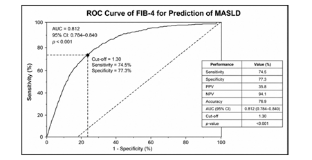
Results: The prevalence of MASLD was 14.2%. Based on the FIB-4 Index, 75.6%, 19.1%, and 5.3% of participants were classified as low-, intermediate-, and high-risk, respectively. Patients with MASLD had significantly higher age, body mass index, duration of diabetes, HbA1c, LDL-C, triglyceride, AST, and ALT levels, and lower HDL-C levels than those without MASLD (p < 0.05). Multivariable logistic regression showed that triglyceride levels ≥150 mg/dL (adjusted OR = 2.08; 95% CI: 1.57–2.75) and intermediate/high FIB-4 risk (adjusted OR = 1.96; 95% CI: 1.42–2.72) were independently associated with MASLD. At a cutoff value of 1.30, the FIB-4 Index demonstrated 74.5% sensitivity, 77.3% specificity, 76.9% accuracy, and an area under the ROC curve (AUC) of 0.812 (95% CI: 0.784–0.840), indicating good discriminative performance for distinguishing patients with and without MASLD.
Conclusion: The prevalence of MASLD among patients with T2DM was 14.2%. Elevated triglyceride levels and intermediate/high FIB-4 risk were independently associated with MASLD. Although the FIB-4 Index is not intended for the diagnosis or screening of MASLD, it demonstrated good discriminative performance and may serve as a practical non-invasive tool for liver fibrosis risk stratification, facilitating the identification of patients who may benefit from further evaluation, such as transient elastography (FibroScan), or referral to a specialist in accordance with current clinical practice guidelines.
References
Younossi ZM. Global epidemiology of metabolic dysfunction-associated steatotic liver disease. Nat Rev Gastroenterol Hepatol. 2024;21(1):40-55.
Lee HH, Kim JH, Park SY. Metabolic dysfunction-associated steatotic liver disease and risk of cardiovascular disease in a nationwide cohort. Gut. 2024;73(2):210-9.
Chung GE, Yu SJ, Yoo JJ, Cho Y, Lee K na, Shin DW, et al. Metabolic dysfunction-associated steatotic liver disease increases cardiovascular disease risk in young adults. Sci Rep. 2025;15(1):5777. doi:10.1038/s41598-025-89293-6.
Chan WK, Chuah KH, Rajaram RB, Lim LL, Ratnasingam J, Vethakkan SR. Metabolic dysfunction-associated steatotic liver disease: a state-of-the-art review. J Obes Metab Syndr. 2023;32(3):197-213. doi: 10.7570/jomes23052.
Targher G, Byrne CD, Tilg H. MASLD: a systemic metabolic disorder with cardiovascular and malignant complications. Gut 2024;73(4):691-702. doi: 10.1136/gutjnl-2023-330595.
Rinella ME, Neuschwander-Tetri BA, Siddiqui MS, Abdelmalek MF, Caldwell S, Barb D, et al. AASLD practice guidance on the clinical assessment and management of nonalcoholic fatty liver disease. Hepatology. 2023;77(5):1797-835. doi: 10.1097/HEP.0000000000000323.
European Association for the Study of the Liver, European Association for the Study of Diabetes, European Association for the Study of Obesity. EASL-EASD-EASO Clinical practice guidelines on the management of metabolic dysfunction-associated steatotic liver disease (MASLD). J Hepatol. 2024;81(3):492-542. doi: 10.1016/j.jhep.2024.04.031.
Sono S. Prevalence and fibrosis status of metabolic dysfunction-associated fatty liver disease in Thailand: a population-based study. Sci Rep. 2025;15(1):1432.
International Diabetes Federation. IDF Diabetes Atlas. 11th ed. Brussels: International Diabetes Federation; 2024.
Ministry of Public Health. Health Data Center (HDC): Non-communicable diseases statistics, Ratchaburi Province, fiscal years 2022-2024. Nonthaburi: Ministry of Public Health; 2024.
American Diabetes Association Professional Practice Committee. Comprehensive medical evaluation and assessment of comorbidities: standards of care in diabetes-2025. Diabetes Care. 2025;48(Suppl 1):S59-S85. doi: 10.2337/dc25-S004.
Downloads
Published
How to Cite
Issue
Section
License
Copyright (c) 2026 Ratchabhiwat Medical Journal

This work is licensed under a Creative Commons Attribution-NonCommercial-NoDerivatives 4.0 International License.
Articles in this journal are copyrighted by the x may be read and used for academic purposes, such as teaching, research, or citation, with proper credit given to the author and the journal.use or modification of the articles is prohibited without permission.
statements expressed in the articles are solely the opinions of the authors.
authors are fully responsible for the content and accuracy of their articles.
other reuse or republication requires permission from the journal."


