
Superior mediastinal cut-off measurements in chest radiographs for the screening of silent thoracic aortic diseases
Keywords:
Chest radiograph, cut-off values, mediastinal measurement, nontraumatic aortic disease, widening of mediastinumAbstract
Background: Analyzing the diagnostic cut-off values of chest radiographs provides an in-depth understanding of unrecognized aortic diseases, thereby contributing to a more comprehensive approach to identifying subtle aortic abnormalities.
Objectives: The objective of this study was to determine the cut-off measurements for superior mediastinal width and mediastinal width-to-thoracic cage ratio from chest radiographs for the early detection of unrecognized thoracic aortic diseases. In addition, this study aimed to enhance diagnostic strategies for identifying aortic abnormalities.
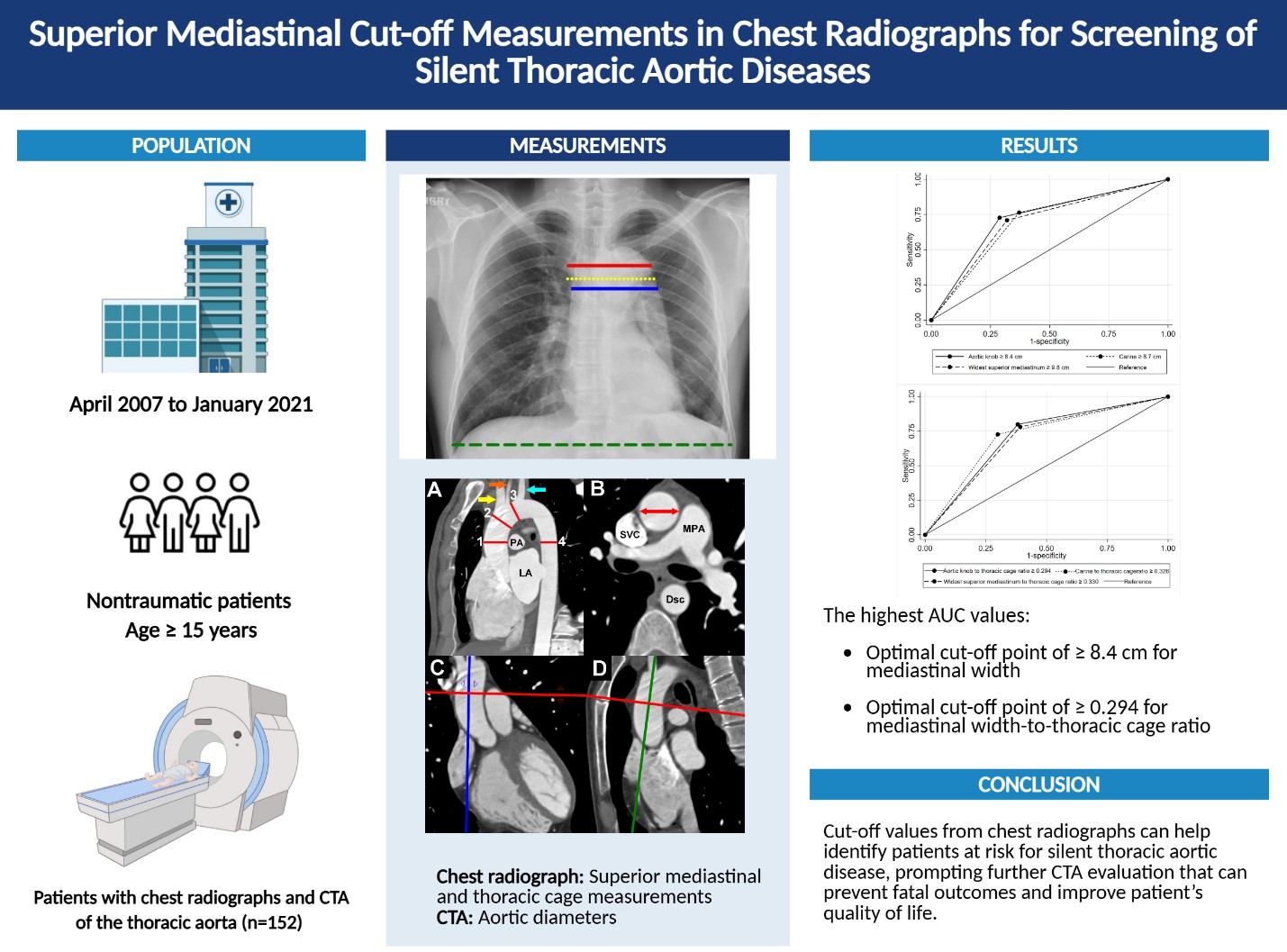
Methods: This retrospective analysis included 152 patients who underwent computed tomography angiography (CTA) of the thoracic aorta and chest radiograph between April 2007 and January 2021. Superior mediastinal width was measured at three levels on chest radiographs. Thoracic CTA findings were categorized into normal and abnormal groups, the latter comprising aortic dissection, intramural hematoma, penetrating atherosclerotic ulcer, and aneurysm. The cut-off values for the superior mediastinal width and mediastinal width-to-thoracic cage ratio were calculated using receiver operating characteristic (ROC) curve analysis.
Results: The optimal cut-off values were a mediastinal width ≥8.4 cm at the aortic knob level (ROC 77.3, 95% confidence interval (CI) 69.8–84.9, P < 0.001; sensitivity 72.7%, specificity 71.1%, positive predictive value (PPV) 58.8%, negative predictive value (NPV) 82.1%) and a mediastinal width-to-thoracic cage ratio ≥ 0.294 at the same level (ROC 78.6, 95% CI 70.8–86.3, P < 0.001; sensitivity 80.0%, specificity 61.9%, PPV 54.3%, NPV 84.5%).
Conclusion: The established superior mediastinal width and ratio cut-off values from chest radiographs can aid clinicians in identifying patients who are at risk for silent thoracic aortic disease, prompting further CTA evaluation that can prevent fatal outcomes and improve patients’ quality of life.
Downloads
References
Holloway BJ, Rosewarne D, Jones RG. Imaging of thoracic aortic disease. Br J Radiol. 2011;84 Spec No 3:S338-S354.
https://doi.org/10.1259/bjr/30655825
Huliyurdurga Srinivasasetty NS, Thagachagere Ramegowda R, Kharge J, Bachahalli Krishnanayak G, S Patil S, Raj V, et al. Unusual non progressive idiopathic giant ascending aortic aneurysm-A rarity. Int J Surg Case Rep 2016;25:203-6.
https://doi.org/10.1016/j.ijscr.2016.06.049
Elefteriades JA, Rizzo JA. Epidemiology prevalence incidence trends. In: Elefteriades J, editor. Acute aortic disease. New York: Informa Healthcare; 2007. p.89-98.
https://doi.org/10.3109/9781420019766
Kälsch H, Lehmann N, Möhlenkamp S, Becker A, Moebus S, Schmermund A, et al. Body-surface adjusted aortic reference diameters for improved identification of patients with thoracic aortic aneurysms: results from the population-based Heinz Nixdorf Recall study. Int J Cardiol 2013;163:72-8.
https://doi.org/10.1016/j.ijcard.2011.05.039
Itani Y, Watanabe S, Masuda Y, Hanamura K, Asakura K, Sone S, et al. Measurement of aortic diameters and detection of asymptomatic aortic aneurysms in a mass screening program using a mobile helical computed tomography unit. Heart Vessels 2002;16:42-5.
https://doi.org/10.1007/s380-002-8315-1
Wongwanit C, Mutirangura P, Chierakul N, Chaiyasoot W, Phongraweewan O. Rapidly enlarging and asymptomatic abdominal aortic aneurysm in a male patient with chronic obstructive pulmonary disease: A case report of endovascular aortic aneurysm repair (EVAR). Siriraj Med J 2006:58;812-8.
Aggarwal S, Qamar A, Sharma V, Sharma A. Abdominal aortic aneurysm: A comprehensive review. Exp Clin Cardiol 2011;16:11-5.
Schermerhorn M. A 66-year-old man with an abdominal aortic aneurysm: review of screening and treatment. JAMA 2009:302;2015-22.
https://doi.org/10.1001/jama.2009.1502
Harris LM, Faggioli GL, Fiedler R, Curl GR, Ricotta JJ. Ruptured abdominal aortic aneurysms: factors affecting mortality rates. J Vasc Surg 1991;14:812-8.
https://doi.org/10.1067/mva.1991.33494
Davies RR, Gallo A, Coady MA, Tellides G, Botta DM, Burke B, et al. Novel measurement of relative aortic size predicts rupture of thoracic aortic aneurysms. Ann Thorac Surg 2006;81:169-77.
https://doi.org/10.1016/j.athoracsur.2005.06.026
Johansson G, Markström U, Swedenborg J. Ruptured thoracic aortic aneurysms: a study of incidence and mortality rates. J Vasc Surg 1995;21:985-8.
https://doi.org/10.1016/S0741-5214(95)70227-X
Prapassaro T, Chinsakchai K, Techarattanaprasert S, Wongwanit C, Ruansetakit C, Hongku K, et al. Determining perioperative mortality in patients with ruptured abdominal aortic aneurysm: Insights from a retrospective cohort study. Siriraj Med J 2024:76;480-7.
https://doi.org/10.33192/smj.v76i8.266315
Asouhidou I, Asteri T. Acute aortic dissection: be aware of misdiagnosis. BMC Res Notes 2009;2:25.
https://doi.org/10.1186/1756-0500-2-25
Chung JH, Ghoshhajra BB, Rojas CA, Dave BR, Abbara S. CT angiography of the thoracic aorta. Radiol Clin North Am 2010;48:249-64.
https://doi.org/10.1016/j.rcl.2010.02.001
McMahon MA, Squirrell CA. Multidetector CT of Aortic Dissection: A Pictorial Review. Radiographics 2010;30:445-60.
https://doi.org/10.1148/rg.302095104
Kapustin AJ, Litt HI. Diagnostic imaging for aortic dissection. Semin Thorac Cardiovasc Surg 2005;17: 214-23.
https://doi.org/10.1053/j.semtcvs.2005.06.006
Nienaber CA, von Kodolitsch Y, Nicolas V, Siglow V, Piepho A, Brockhoff C, et al. The diagnosis of thoracic aortic dissection by noninvasive imaging procedures.N Engl J Med 1993;328:1-9.
https://doi.org/10.1056/NEJM199301073280101
Mao SS, Ahmadi N, Shah B, Beckmann D, Chen A, Ngo L, et al. Normal thoracic aorta diameter on cardiac computed tomography in healthy asymptomatic adults: impact of age and gender. Acad Radiol 2008;15: 827-34.
https://doi.org/10.1016/j.acra.2008.02.001
Stein E, Mueller GC, Sundaram B. Thoracic aorta (multidetector computed tomography and magnetic resonance evaluation). Radiol Clin North Am 2014;52:195-217.
https://doi.org/10.1016/j.rcl.2013.08.002
Kimura-Hayama ET, Meléndez G, Mendizábal AL, Meave-González A, Zambrana GFB, Corona-Villalobos CP. Uncommon congenital and acquired aortic diseases: role of multidetector CT angiography. Radiographics 2010;30:79-98.
https://doi.org/10.1148/rg.301095061
Liotta R, Chughtai A, Agarwal PP. Computed tomography angiography of thoracic aortic aneurysms. Semin Ultrasound CT MR 2012;33:235-46.
https://doi.org/10.1053/j.sult.2011.11.003
Prabhasavat, K. Measurement of the aortic diameter in the asymptomatic Thai population in Siriraj Hospital: Assessment with multidetector CT. Siriraj Med J 2016;68: 247-56.
Siriapisith T, Tongdee T, Tongdee R, Jitnuson P, Karuvanarint S. CT criteria for differentiation between true and false lumen in aortic dissection. Siriraj Med J 2004;56:410-17.
Kooiman J, Pasha SM, Zondag W, Sijpkens YWJ, van der Molen AJ, Huisman MV, et al. Meta-analysis: serum creatinine changes following contrast enhanced CT imaging. Eur J Radiol 2012;81:2554-61.
https://doi.org/10.1016/j.ejrad.2011.11.020
Solomon R. Contrast-medium-induced acute renal failure. Kidney Int 1998;53:230-42.
https://doi.org/10.1038/sj.ki.4495510
Morcos SK, Thomsen HS, Webb JA. Contrast-mediainduced nephrotoxicity: a consensus report. Contrast Media Safety Committee, European Society of Urogenital Radiology (ESUR). Eur Radiol 1999;9:1602-13.
https://doi.org/10.1007/s003300050894
Rudnick MR, Goldfarb S, Wexler L, Ludbrook PA, Murphy MJ, Halpern EF, et al. Nephrotoxicity of ionic and nonionic contrast media in 1196 patients: a randomized trial. The Iohexol Cooperative Study. Kidney Int 1995;47:254-61.
https://doi.org/10.1038/ki.1995.32
Pistolesi V, Regolisti G, Morabito S, Gandolfini I, Corrado S, Piotti G, et al. Contrast medium induced acute kidney injury: a narrative review. J Nephrol 2018;31:797-812.
https://doi.org/10.1007/s40620-018-0498-y
Karlsberg RP, Dohad SY, Sheng R. Contrastinduced acute kidney injury (CI-AKI) following intra-arterial administration of iodinated contrast media. J Nephrol 2010;23:658-66.
https://doi.org/10.1093/joneph/23.6.658
Karlsberg RP, Dohad SY, Sheng R. Contrast medium-induced acute kidney injury: comparison of intravenous and intraarterial administration of iodinated contrast medium. J Vasc Interv Radiol2011;22:1159-65.
https://doi.org/10.1016/j.jvir.2011.03.020
Mohammed NM, Mahfouz A, Achkar K, Rafie IM, Hajar R. Contrast-induced Nephropathy. Heart Views 2013;14:106-16.
https://doi.org/10.4103/1995-705X.125926
Choorat S, Totanarungroj K, Muangman, N. Assessment of normal subcarinal angle on chest radiographs in adult Thai population. Siriraj Med J 2008;60:264-6.
Somcharit L. Traumatic Hemothorax and Pneumothorax detected by EFAST Compared with Chest Radiography at Siriraj Hospital. Siriraj Med J 2016;68:171-6.
Hiratzka LF, Bakris GL, Beckman JA, et al. 2010. ACCF/AHA/AATS/ACR/ASA/SCA/SCAI/SIR/STS/SVM guidelines for the diagnosis and management of patients with Thoracic Aortic Disease: a report of the American College of Cardiology Foundation/American Heart Association Task Force on Practice Guidelines, American Association for Thoracic Surgery, American College of Radiology, American Stroke Association, Society of Cardiovascular Anesthesiologists, Society for Cardiovascular Angiography and Interventions, Society of Interventional Radiology, Society of Thoracic Surgeons, and Society for Vascular Medicine. Circulation 2010;122:e410.
Riambau V, Böckler D, Brunkwall J, Cao P, Chiesa R, Coppi G, et al. Editor's choice - management of descending thoracic aorta diseases: clinical practice guidelines of the European Society for Vascular Surgery (ESVS). Eur J Vasc Endovasc Surg 2017;53:4-52.
https://doi.org/10.1016/j.ejvs.2016.06.005
Fultz PJ, Melville D, Ekanej A, Holzwasser G, Voci S, Wandtke JC, et al. Nontraumatic rupture of the thoracic aorta: chest radiographic features of an often unrecognized condition. AJR Am J Roentgenol 1998;171:351-7.
https://doi.org/10.2214/ajr.171.2.9694450
Seltzer SE, D'Orsi C, Kirshner R, DeWeese JA. Traumatic aortic rupture: plain radiographic findings. AJR Am J Roentgenol 1981;137:1011-4.
https://doi.org/10.2214/ajr.137.5.1011
Marnocha KE, Maglinte DD, Woods J, Goodman M, Peterson P. Mediastinal-width/chest-width ratio in blunt chest trauma: a reappraisal. AJR Am J Roentgenol 1984;142:275-7.
https://doi.org/10.2214/ajr.142.2.275
Pham MHC, Sigvardsen PE, Fuchs A, Kühl JT, Sillesen H, Afzal S, et al. Aortic aneurysms in a general population cohort: prevalence and risk factors in men and women. Eur Heart J Cardiovasc Imaging 2024;25:1235-43.
Downloads
Published
How to Cite
Issue
Section
License
Copyright (c) 2026 Chulalongkorn Medical Journal

This work is licensed under a Creative Commons Attribution-NonCommercial-NoDerivatives 4.0 International License.


