
Modified DCER technique for Basilar invagination and atlantoaxial dislocation in neurofibromatosis type 1: A Case Report
คำสำคัญ:
Neurofibromatosis Type 1 (NF-1), Atlantoaxial Dislocation (AAD), Basilar Invagination (BI), DCER technique, Craniovertebral Junction Anomaliesบทคัดย่อ
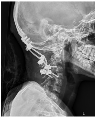
Introduction: Spinal deformities are common in Neurofibromatosis Type 1 (NF-1), a rare autosomal dominant disorder, however, craniovertebral junction anomalies such as atlantoaxial dislocation (AAD) and basilar invagination (BI) are uncommon. Surgical management is indicated in symptomatic cases. Although combined anterior–posterior fixation remains the standard approach, the distraction, compression, and extension reduction (DCER) technique offers a safe and effective alternative in selected patients. Case presentation: A 21-year-old woman with clinical features of Neurofibromatosis Type 1 (NF-1) presented with sudden-onset quadriparesis following a fall to the ground. Neuroimaging revealed atlantoaxial dislocation (AAD) and basilar invagination (BI). She was initially managed with cervical traction, followed by a single-stage posterior fixation and fusion using a modified distraction, compression, and extension reduction (DCER) technique. Postoperatively, spinal alignment and stability were restored, and the patient demonstrated marked neurological improvement. Conclusion: Surgical management of atlantoaxial dislocation (AAD) and basilar invagination (BI) in Neurofibromatosis Type 1 (NF-1) is particularly challenging due to the presence of dystrophic anatomy. The posterior-only distraction, compression, and extension reduction (DCER) technique offers an effective, patient-specific approach for deformity correction and achieving optimal outcomes.
Downloads
เอกสารอ้างอิง
Legius E, Messiaen L, Wolkenstein P, Pancza P, Avery RA, Berman Y, et al. Revised diagnostic criteria for neurofibromatosis type 1 and Legius syndrome: an international consensus recommendation. Genet Med 2021;23(8):1506-13. https://doi.org/10.1038/s41436-021-01170-5
Well L, Careddu A, Stark M, Farschtschi S, Bannas P, Adam G, et al. Phenotyping spinal abnormalities in patients with Neurofibromatosis type 1 using whole-body MRI. Sci Rep. 2021;11(1):16889. https://doi.org/10.1038/s41598-021-96310-x
Ding C, Guo Y, Wu T, Wang B, Huang K, He J, et al. Atlantoaxial Dislocation Associated with Type 1 Neurofibromatosis: Case Report and Review of the Literature. World Neurosurg. 2020;143:261-7. https://doi.org/10.1016/j.wneu.2020.07.176
Yang SY, Boniello AJ, Poorman CE, Chang AL, Wang S, Passias PG. A review of the diagnosis and treatment of atlantoaxial dislocations. Global Spine J. 2014;4(3):197-210. https://doi.org/10.1055/s-0034-1376371
Chandra PS, Bajaj J, Singh PK, Garg K, Agarwal D. Basilar Invagination and Atlantoaxial Dislocation: Reduction, Deformity Correction and Realignment Using the DCER (Distraction, Compression, Extension, and Reduction) Technique With Customized Instrumentation and Implants. Neurospine. 2019;16(2):231-50. https://doi.org/10.14245/ns.1938194.097
Kossler N, Stricker S, Rödelsperger C, Robinson PN, Kim J, Dietrich C, et al. Neurofibromin (Nf1) is required for skeletal muscle development. Hum Mol Genet. 2011;20(14):2697-709. https://doi.org/10.1093/hmg/ddr149
Yang SY, Boniello AJ, Poorman CE, Chang AL, Wang S, Passias PG. A review of the diagnosis and treatment of atlantoaxial dislocations. Global Spine J. 2014;4(3):197-210. https://doi.org/10.1055/s-0034-1376371
Chandra PS, Kumar A, Chauhan A, Ansari A, Mishra NK, Sharma BS. Distraction, compression, and extension reduction of basilar invagination and atlantoaxial dislocation: a novel pilot technique. Neurosurgery. 2013;72(6):1040-53.
https://doi.org/10.1227/NEU.0b013e31828bf342
Chandra PS, Prabhu M, Goyal N, Garg A, Chauhan A, Sharma BS. Distraction, Compression, Extension, and Reduction Combined with Joint Remodeling and Extra-articular Distraction: Description of 2 New Modifications for Its Application in Basilar Invagination and Atlantoaxial Dislocation: Prospective Study in 79 Cases. Neurosurgery. 2015;77(1):67-80. doi:10.1227/ NEU.0000000000000737
ดาวน์โหลด
เผยแพร่แล้ว
รูปแบบการอ้างอิง
ฉบับ
ประเภทบทความ
สัญญาอนุญาต
ลิขสิทธิ์ (c) 2026 วารสารประสาทศัลยศาสตร์ไทย

อนุญาตภายใต้เงื่อนไข Creative Commons Attribution-NonCommercial-NoDerivatives 4.0 International License.
##default.contextSettings.thaijo.licenseTerms##

