
รายงานผู้ป่วยเนื้องอกสมองชนิดกลัยโอมาระดับรุนแรงในทารก จากภาคใต้ของประเทศไทย
คำสำคัญ:
Infantile high-grade glioma, neuro-oncology, seizure, glioma, infant-type hemispheric gliomasบทคัดย่อ
บทนำ: เนื้องอกสมองชนิดกลัยโอมาระดับรุนแรง (High-grade glioma; HGG) ในทารกอายุต่ำกว่า 1 ปี เป็นโรคที่พบได้น้อยและมีลักษณะทางชีววิทยาแตกต่างจากกลัยโอมาระดับรุนแรงในเด็กโตและผู้ใหญ่ อาการแสดงมักไม่จำเพาะและค่อยเป็นค่อยไป ทำให้การวินิจฉัยในระยะเริ่มต้นเป็นไปได้ยาก โดยเฉพาะในบริบทของประเทศที่มีทรัพยากรจำกัด
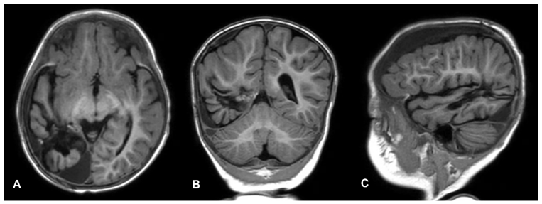
รายงานผู้ป่วย: รายงานผู้ป่วยเด็กหญิงไทยอายุ 11 เดือน มีอาการอาเจียนตอนเช้าเพิ่มขึ้นต่อเนื่องเป็นเวลา 1 เดือน ร่วมกับอาการหงุดหงิด จับศีรษะบ่อย และหกล้มซ้ำ ๆ ก่อนมาโรงพยาบาลเกิดอาการชักชนิด generalized tonic-clonic การตรวจด้วยคลื่นแม่เหล็กไฟฟ้าสมองพบก้อนเนื้องอกขนาดใหญ่เหนือ tentorium ร่วมกับภาวะกดเบียดโครงสร้างสมอง ผู้ป่วยได้รับการผ่าตัดเอาก้อนเนื้องอกออกทั้งหมด การตรวจทางพยาธิวิทยาพบลักษณะของกลิโอมาระดับรุนแรง มีเซลล์ผิดรูป การแบ่งตัวสูง การเพิ่มจำนวนของหลอดเลือด และค่า Ki-67 ร้อยละ 35 การย้อมภูมิคุ้มกันพบ GFAP ให้ผลบวก H3K27me3 ให้ผลบวก และ IDH1-R132H ให้ผลลบ ไม่สามารถตรวจการกลายพันธุ์แบบ gene fusion ได้ หลังผ่าตัดผู้ป่วยฟื้นตัวดี และมีแผนให้เคมีบำบัดตามแนวทาง Thai POG-BT-131FB
อภิปราย: กลัยโอมาระดับรุนแรงในทารกเป็นโรคที่พบได้น้อยและมีลักษณะเฉพาะทางชีววิทยา อาการไม่จำเพาะอาจทำให้การวินิจฉัยล่าช้า โดยอาการอาเจียนตอนเช้าอย่างต่อเนื่องและอาการชักเฉียบพลันถือเป็นสัญญาณเตือนสำคัญ การผ่าตัดเอาก้อนเนื้องอกออกอย่างปลอดภัยมากที่สุดเป็นหัวใจสำคัญของการรักษา เนื่องจากการฉายรังสีในทารกมีความเสี่ยงสูงต่อพัฒนาการทางระบบประสาท จึงนิยมใช้เคมีบำบัดเป็นการรักษาเสริมระยะแรก
สรุป: รายงานผู้ป่วยนี้เน้นย้ำถึงเบาะแสสำคัญในการวินิจฉัยกลัยโอมาระดับรุนแรงในทารก และความจำเป็นของการตรวจภาพสมองตั้งแต่ระยะแรกในเด็กที่มีอาการอาเจียนเรื้อรังหรืออาการทางระบบประสาทเฉียบพลัน การรักษาแบบสหสาขา รวมถึงการผ่าตัดและเคมีบำบัด มีบทบาทสำคัญในการดูแลผู้ป่วยกลุ่มนี้
Downloads
เอกสารอ้างอิง
Ostrom QT, de Blank PM, Kruchko C, et al. Alex’s Lemonade Stand Foundation infant and childhood primary brain and central nervous system tumors diagnosed in the United States in 2007–2011. Neuro Oncol. 2014;16(Suppl 10):x1–36. doi:10.1093/neuonc/nou327.
Fangusaro J. Pediatric high grade glioma: a review and update on tumor clinical characteristics and biology. Front Oncol. 2012;2:105. doi:10.3389/fonc.2012.00105.
Louis DN, Perry A, Wesseling P, Brat DJ, Cree IA, Figarella-Branger D, et al. The 2021 WHO classification of tumors of the central nervous system: a summary. Neuro Oncol. 2021;23(8):1231–51. doi:10.1093/neuonc/noab106.
Chavaz L, Bagchi A, Dhanda SK, et al. A systematic study of molecular diagnosis, treatment and prognosis in infant-type hemispheric glioma: an individual patient data meta-analysis of 164 patients. Neuro Oncol. 2025 Nov 8;noaf264. Epub ahead of print. doi:10.1093/neuonc/noaf264. PMID:41206756.
Bagchi A, Chiang J, Pinto S, Dhanda S, Gajjar A. Infant-type hemispheric gliomas: a review of clinical, radiologic, histopathologic, and molecular features. J Natl Compr Canc Netw. 2025;23(11):e257064. doi:10.6004/jnccn.2025.7064.
Guerreiro Stucklin AS, Ryall S, Fukuoka K, Zapotocky M, Lassaletta A, Li C, Bridge T, Kim B, et al. Alterations in ALK/ROS1/NTRK/MET drive a group of infantile hemispheric gliomas. Nat Commun. 2019;10(1):4343. doi:10.1038/s41467-019-12187-5.
Buccoliero AM, Giunti L, Moscardi S, Castiglione F, Provenzano A, Sardi I, et al. Pediatric high-grade glioma classification criteria and molecular features of a case series. Genes (Basel). 2022;13(4):624. doi:10.3390/genes13040624.
Jones C, Karajannis MA, Jones DTW, Kieran MW, Monje M, Baker SJ, et al. Pediatric high-grade glioma: biologically and clinically in need of new thinking. Neuro Oncol. 2017;19(2):153–61. doi:10.1093/neuonc/now101.
Baugh JN, Gielen GH, van Vuurden DG, Veldhuijzen van Zanten SEM, Hargrave D, Massimino M, et al. Transitioning to molecular diagnostics in pediatric high-grade glioma: experiences with the 2016 WHO classification of CNS tumors. Neurooncol Adv. 2021;3(1):vdab113. doi:10.1093/noajnl/vdab113.
Papusha L, Zaytseva M, Senchenko M, Panferova A, Sanakoeva A, Artemov A, et al. Challenges in diagnostics and treatment of infant-type hemispheric gliomas. Neurooncol Adv. 2025;7(1):vdaf124. doi:10.1093/noajnl/vdaf124.
Lu VM, O’Connor KP, Himes BT, Brown DA, Nesvick CL, Siada RG, et al. Effect of surgery and chemotherapy on long-term survival in infants with congenital glioblastoma: an integrated survival analysis. J Neurosurg Pediatr. 2020;26(5):563–71. doi:10.3171/2020.5.PEDS20226.
Tunthanathip T, Madteng S. Factors associated with the extent of resection of glioblastoma. Precis Cancer Med. 2020;3:12. doi:10.21037/pcm.2020.01.01.
Major N, Patel NA, Bennett J, Novakovic E, Poloni D, Abraham M, et al. The current state of radiotherapy for pediatric brain tumors: an overview of post-radiotherapy neurocognitive decline and outcomes. J Pers Med. 2022;12(7):1050. doi:10.3390/jpm12071050.
Chiang J, Bagchi A, Li X, Dhanda SK, Huang J, Pinto SN, et al. High-grade glioma in infants and young children is histologically, molecularly, and clinically diverse: results from the SJYC07 trial and institutional experience. Neuro Oncol. 2024;26(1):178–90. doi:10.1093/neuonc/noad130.
Tunthanathip T, Sae-Heng S, Oearsakul T, Kaewborisutsakul A, Taweesomboonyat C. Economic impact of a machine learning-based strategy for preparation of blood products in brain tumor surgery. PLoS One. 2022;17(7):e0270916. doi:10.1371/journal.pone.0270916.
Clarke M, Mackay A, Ismer B, Pickles JC, Tatevossian RG, Newman S, et al. Infant high-grade gliomas comprise multiple subgroups characterized by novel targetable gene fusions and favorable outcomes. Cancer Discov. 2020;10(7):942–63. doi:10.1158/2159-8290.CD-19-1030.
Jitchanvichai J, Tunthanathip T. Cost-effectiveness of intracranial pressure monitoring in severe traumatic brain injury in Southern Thailand. Acute Crit Care. 2025;40(1):69–78. doi:10.4266/acc.004080.
ดาวน์โหลด
เผยแพร่แล้ว
รูปแบบการอ้างอิง
ฉบับ
ประเภทบทความ
สัญญาอนุญาต
ลิขสิทธิ์ (c) 2026 วารสารประสาทศัลยศาสตร์ไทย

อนุญาตภายใต้เงื่อนไข Creative Commons Attribution-NonCommercial-NoDerivatives 4.0 International License.
##default.contextSettings.thaijo.licenseTerms##
