Conventional flexible bronchoscopy versus radial endobronchial ultrasound (R-EBUS)-guided bronchoscopy under fluoroscopy: A retrospective study at Phrapokklao Hospital, Chanthaburi, Thailand
Keywords:
Bronchoscopy, diagnostic yield, fluoroscopy, peripheral pulmonary lesion, radial endobronchial ultrasoundAbstract
Background: Conventional flexible bronchoscopy is widely used for evaluating peripheral pulmonary lesions; however, its diagnostic yield remains limited. Although radial endobronchial ultrasound (R-EBUS) combined with fluoroscopic guidance may improve lesion localization and tissue acquisition, real-world comparative data from provincial tertiary-care centers are scarce.
Objectives: To compare the diagnostic yield of conventional flexible bronchoscopy and R-EBUS–guided bronchoscopy under fluoroscopic guidance for peripheral pulmonary lesions and identify patient-, lesion-, and procedure-related factors associated with diagnostic success.
Methods: We conducted a retrospective observational study at a provincial tertiary-care hospital in Eastern Thailand. Adult patients undergoing diagnostic bronchoscopy for peripheral pulmonary lesions between October 2023 and October 2025 were included. Diagnostic yield was defined according to contemporary ATS/ ACCP Delphi consensus criteria. Univariable and multivariable logistic regression analyses were performed to evaluate factors associated with diagnostic yield, and Firth penalized logistic regression used as confirmatory analysis to address potential sparse-data bias.
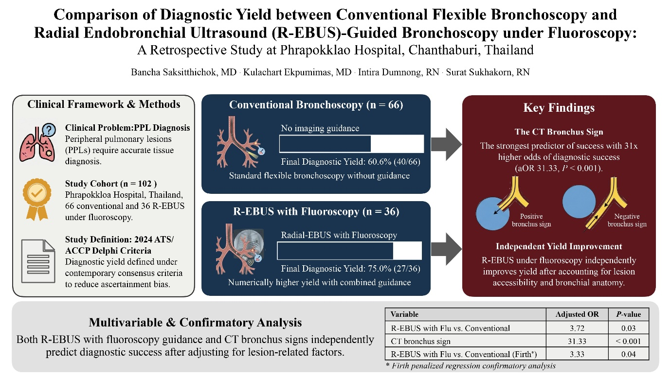
Results: A total of 102 patients were analyzed, including 66 who underwent conventional bronchoscopy and 36 who underwent R-EBUS–guided bronchoscopy under fluoroscopic guidance. The final procedure-level diagnostic yield was higher, but not significantly different, in the R-EBUS with fluoroscopy group than in the conventional group (75.0% vs. 60.6%; P = 0.14). After adjusting for clinically relevant lesion-related factors, REBUS–guided bronchoscopy under fluoroscopic guidance was independently associated with higher diagnostic yield (adjusted odds ratio [OR] 3.72, 95% confidence interval [CI] 1.12–12.38, P = 0.03). Presence of a computed tomography (CT) bronchus sign was the strongest, most consistent predictor of diagnostic success across all models (adjusted OR 31.33, 95% CI 8.43–116.52, P < 0.001). Confirmatory Firth penalized regression yielded consistent estimates in direction and magnitude.
Conclusion: In this real-world provincial tertiary-care setting, R-EBUS–guided bronchoscopy under fluoroscopic guidance was independently associated with higher diagnostic yield after accounting for lesion accessibility, particularly presence of a CT bronchus sign. Lesion-related factors, especially bronchial anatomy, play a central role in determining diagnostic success and should be carefully assessed when selecting bronchoscopic techniques in resource-limited settings.
Downloads
References
Bray F, Laversanne M, Sung H, Ferlay J, Siegel RL, Soerjomataram I, et al. Global cancer statistics 2022: GLOBOCAN estimates of incidence and mortality worldwide for 36 cancers in 185 countries. CA Cancer J Clin 2024;74:229-63.
https://doi.org/10.3322/caac.21834
Mazzone PJ, Silvestri GA, Souter LH, Caverly TJ, Kanne JP, Katki HA, et al. Screening for lung cancer: CHEST guideline and expert panel report Chest 2021;160:e427-94.
https://doi.org/10.1016/j.chest.2021.06.063
Rivera MP, Mehta AC, Wahidi MM. Establishing the diagnosis of lung cancer: Diagnosis and management of lung cancer, 3rd ed: American College of Chest Physicians evidence-based clinical practice guidelines. Chest 2013;143 5 Suppl:e142S-e165S.
https://doi.org/10.1378/chest.12-2353
Baaklini WA, Reinoso MA, Gorin AB, Sharafkaneh A, Manian P. Diagnostic yield of fiberoptic bronchoscopy in evaluating solitary pulmonary nodules. Chest 2000;117:1049-54.
https://doi.org/10.1378/chest.117.4.1049
Ost DE, Ernst A, Lei X, Kovitz KL, Benzaquen S, Diaz-Mendoza J, et al. Diagnostic yield and complications of bronchoscopy for peripheral lung lesions. Results of the AQuIRE registry. Am J Respir Crit Care Med 2016;193:68-77.
https://doi.org/10.1164/rccm.201507-1332OC
Tanner NT, Yarmus L, Chen A, Wang Memoli J, Mehta HJ, Pastis NJ, et al. Standard bronchoscopy with fluoroscopy vs. thin bronchoscopy and radial endobronchial ultrasound for biopsy of pulmonary lesions: A multicenter, prospective, randomized trial. Chest 2018;154:1035-43.
https://doi.org/10.1016/j.chest.2018.08.1026
Gasparini S, Mei F, Bonifazi M, Zuccatosta L. Bronchoscopic diagnosis of peripheral lung lesions. Curr Opin Pulm Med 2022;28:31-6.
https://doi.org/10.1097/MCP.0000000000000842
Hürter T, Hanrath P. Endobronchial sonography: feasibility and preliminary results. Thorax 1992;47:565-7.
https://doi.org/10.1136/thx.47.7.565
Goldberg BB, Steiner RM, Liu JB, Merton DA, Articolo G, Cohn JR, et al. US-assisted bronchoscopy with use of miniature transducer-containing catheters. Radiology 1994;190:233-7.
https://doi.org/10.1148/radiology.190.1.8259411
Balasubramanian P, Abia-Trujillo D, Barrios-Ruiz A, Garza-Salas A, Koratala A, Chandra NC, et al. Diagnostic yield and safety of diagnostic techniques for pulmonary lesions: systematic review, meta-analysis and network meta-analysis. Eur Respir Rev 2024;33:240046.
https://doi.org/10.1183/16000617.0046-2024
Boonsarngsuk V, Raweelert P, Juthakarn S. Endobronchial ultrasound plus fluoroscopy versus fluoroscopy-guided bronchoscopy: a comparison of diagnostic yields in peripheral pulmonary lesions. Lung 2012;190:233-7.
https://doi.org/10.1007/s00408-011-9359-3
Sánchez-Font A, Giralt L, Vollmer I, Pijuan L, Gea J, Curull V. Endobronchial ultrasound for the diagnosis of peripheral pulmonary lesions. A controlled study with fluoroscopy. Arch Bronconeumol 2014;50:166-71.
https://doi.org/10.1016/j.arbres.2013.11.019
Sainz Zuñiga PV, Vakil E, Molina S, Bassett RL Jr, Ost DE. Sensitivity of radial endobronchial ultrasound-guided bronchoscopy for lung cancer in patients with peripheral pulmonary lesions: An updated meta-analysis. Chest 2020;157:994-1011.
https://doi.org/10.1016/j.chest.2019.10.042
Casutt A, Prella M, Beigelman-Aubry C. Fluoroscopic-Guided Radial Endobronchial Ultrasound Without Guide Sheath For Peripheral Pulmonary Lesions: A Safe And Efficient Combination. Arch Bronconeumol 2015;51:338-43.
https://doi.org/10.1016/j.arbres.2014.09.017
Gonzalez AV, Silvestri GA, Korevaar DA, Gesthalter YB, Almeida ND, Chen A, et al. Assessment of advanced diagnostic bronchoscopy outcomes for peripheral lung lesions: A Delphi consensus definition of diagnostic yield and recommendations for patient-centered study designs. An official American Thoracic Society/American College of Chest Physicians research statement. Am J Respir Crit Care Med 2024;209:634-46.
https://doi.org/10.1164/rccm.202401-0192ST
Bezel P, Tischler V, Robinson C, Baumueller S, Bode-Lesniewska B, Kohler M, et al. Diagnostic value of bronchoalveolar lavage for diagnosis of suspected peripheral lung cancer. Clin Lung Cancer 2016;17:e151-6.
https://doi.org/10.1016/j.cllc.2015.12.012
Minezawa T, Okamura T, Yatsuya H, Yamamoto N, Morikawa S, Yamaguchi T, et al. Bronchus sign on thin-section computed tomography is a powerful predictive factor for successful transbronchial biopsy using endobronchial ultrasound with a guide sheath for small peripheral lung lesions: a retrospective observational study. BMC Med Imaging 2015;15:21.
https://doi.org/10.1186/s12880-015-0060-5
Tsuboi E, Ikeda S, Tajima M, Shimosato Y, Ishikawa S. Transbronchial biopsy smear for diagnosis of peripheral pulmonary carcinomas. Cancer 1967;20:687-98.
https://doi.org/10.1002/1097-0142(1967)20:5<687::AID-CNCR2820200521>3.0.CO;2-K
Yuan M, Hu Y, Wang L, Yin W, Xiao Y. Diagnostic outcomes of radial endobronchial ultrasound bronchoscopy guided by manual navigation in the evaluation of peripheral pulmonary lesions: An observational study. Clin Respir J 2024;18:e13768.
Downloads
Published
How to Cite
Issue
Section
License
Copyright (c) 2026 Chulalongkorn Medical Journal

This work is licensed under a Creative Commons Attribution-NonCommercial-NoDerivatives 4.0 International License.